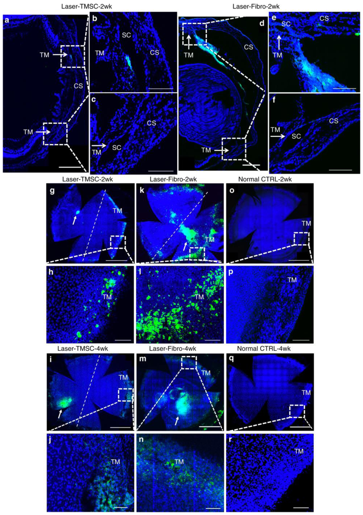
FIGURE 3. Human trabecular meshwork stem cells (TMSCs) home to laser-damaged trabecular meshwork (TM) regions after intracameral injection.

TMSCs and fibroblasts were prelabeled with DiO (green) prior to intracameral injection, following which sections and wholemounts were stained with DAPI (blue). Cryosections (a–f) show localization of injected TMSCs (a–c) or fibroblasts (d–f) 2 weeks after transplantation. Scale bars, 300 μm. b, c, e, f are magnifications of the boxed regions in a, e. b, e show laser-damaged regions, while c, f are unlasered regions. Arrows point to the TM. Scale bars, 100 μm. Wholemounts show distribution of transplanted TMSCs at 2 weeks (g, h) and 4 weeks (i, j), transplanted fibroblasts at 2 weeks (k, l) and 4 weeks (m, n), control eyes without cell transplantation (o–r). The right side of the dotted line is the laser-damaged region, whereas the left side is unlasered region. h, f, p, j, n, r are magnifications of the boxed regions in g, k, o, i, m, q. The green cell clusters on the corneas (white arrows in g, l, k, m) were injected cells healing corneal wounds caused by injection needles. Scale bars, 1 mm (g, k, o, i, m, q); 100 μm (h, f, p, j, n, r). Abbreviations: SC Schlemm’s canal, CS corneal stroma. Adapted from Yun, H., et al. Commun Biol 2018; 1(1): 216 under the Creative Commons Attribution 4.0 International (CCBY4.0) license.
