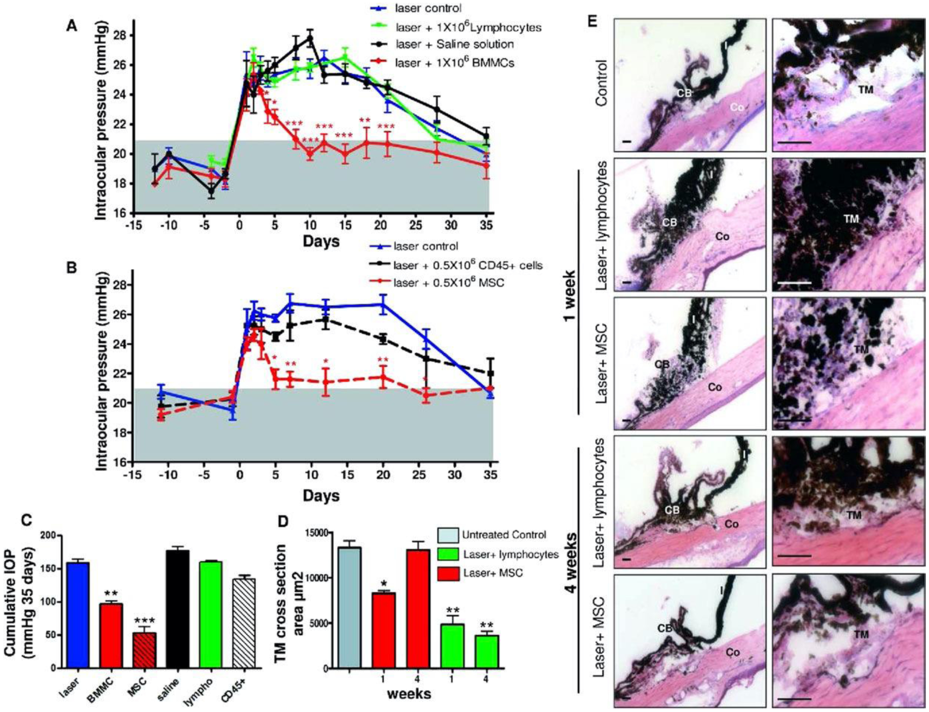
FIGURE 5. Mesenchymal stem cells (MSCs) induce a rapid return to normal intraocular pressure (IOP) levels in experimental glaucoma.

(A) Anterior chambers were injected either with 1 × 10⁶ bone marrow-derived mesenchymal cells (BMMC, red), 1 × 10⁶ lymphocytes (black), saline (green), or received no additional treatment (blue). The gray area represents IOP normal range. IOP was reported as mean ± SEM of four experiments (12 animals per group). (B) 0.5 × 10⁶ CD45+ cells (black) or 0.5 × 10⁶ MSC (red line) were injected intraocularly after laser exposure and IOP was evaluated as described above. We plot mean ± SEM of three experiments (9 animals per group). (C) Cumulative IOP exposure in eyes that received laser damage and injection of different cellular populations and controls. Cumulative IOP exposure is calculated as the temporal integral of IOP over the 4-week experimental period. (D) TM cross-sectional area from histologic sections for each experimental group (red, laser þ MSC group; green, laser þ lymphocyte group) and for a naïve TM (gray) (E) Representative images of hematoxylin-eosin (H&E) stained rat ocular anterior segments before (C: control) and 1 week after laser exposure alone or followed by MSC injection, as well as 4 weeks after laser damage alone or followed by MSC injection. Significance compared to laser control group: *, p<0.05; **, p<0.01; ***, p<0.001. Scale bar: 450μm. Abbreviations: CB, ciliary body; Co, cornea; I, iris. Reproduced with permission from Manuguerra-GagnÉ et al. Stem Cells. 2013;31:1136–1148. (license 1139632-2)
