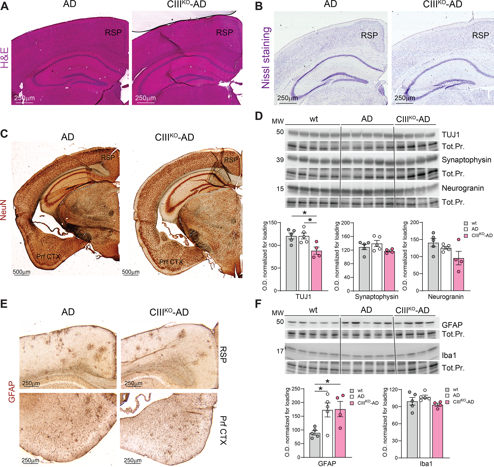
Figure4: Neuronal content and Neuroinflammation:

(A) Representative images of H&E staining on histological sections from AD and CIIIKO-AD mice. (B) Representative images of Nissl staining on histological sections from AD and CIIIKO-AD mice. (C) Representative images of IHC probing for neuronal marker NeuN staining on coronal sections of 8-month-old AD and CIIIKO-AD females. (D) Western blot probing for Class III β-Tubulin (TUJ1), Synaptophysin, and Neurogranin on cortical homogenates from 8-month-old WT, AD, and CIIIKO-AD females (n=4–5/group) and relative quantifications normalized to loading. (E) Representative images of IHC probing for glial marker GFAP on cortex of 8-month-old AD and CIIIKO-AD females. (F) Western blot probing for glial marker GFAP and microglial marker Iba1 on cortical homogenates from 8-month-old WT, AD, and CIIIKO-AD females (n=4–5/group) and relative quantifications normalized to loading. RSP: Retrosplenial area; Prf CTX: Piriform Cortex. Tot.Pr.: Total Protein.
