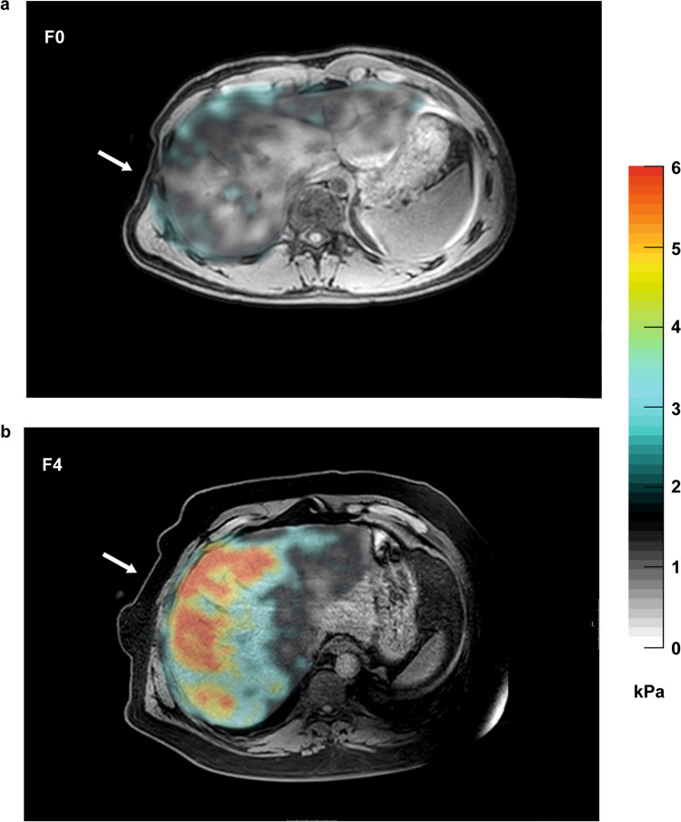
Fig. 3.
Hepatic magnetic resonance elastography (MRE) images captured from two NAFLD patients using an active electrodynamic transducer transmitting mechanical waves at 54 Hz. Arrow indicates placement of transducer when strapped to the patient. Tissue stiffness is illustrated in a corresponding elastogram, shown as a color gradient with higher degrees of tissue stiffness indicated in red. Liver biopsies obtained immediately after the MRE-examination showed a absence of fibrosis (F0) and b cirrhosis (F4), respectively. Abbreviations: kPa, kilopascal; NAFLD, non-alcoholic fatty liver disease

