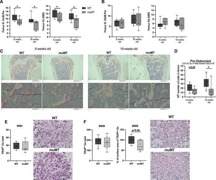
Fig. 4.

Reduced number of osteoclasts in mice lacking mature B cells and immunoglobulins. (A) The N.Oc/B.Pm and Oc.S/BS in the distal metaphyseal part of femur of 6‐week‐old and 16‐week‐old female muMT and WT littermate mice. Student's t test was used to assess differences between WT and muMT mice. n = 6–10. *p < 0.05. (B) The N.Ob/B.Pm and Ob.S/BS in the distal metaphyseal part of femur of 6‐week‐old and 16‐week‐old female muMT and WT littermate mice. Student's t test was used to assess the differences between WT and muMT mice. n = 6–10. (C) Representative images of TRAP‐positive osteoclasts in the femur. (D) The number of pre‐osteoclasts (CD11b+, Gr1−, F480+, RANK+, MCSF‐R+) in bone marrow of femur in 6‐week‐old and 16‐week‐old female muMT and WT mice. Student's t test was used to assess differences between WT and muMT mice at each time point. n = 8–10. *p < 0.05. Ex vivo RANKL stimulated osteoclast differentiation cultures from (E) crude BMC, n = 6, and (F) BMM, n = 8–9. Student's t test was used to assess the differences between WT and muMT mice. BMC = bone marrow cells; BMM = bone marrow macrophages; N.Ob/B.Pm = number of osteoblasts per bone perimeter; N.Oc/B.Pm = number of osteoclasts per bone perimeter; Ob.S/BS = osteoblast surface per bone surface; Oc.S/BS = osteoclast surface per bone surface; WT = wild‐type.
