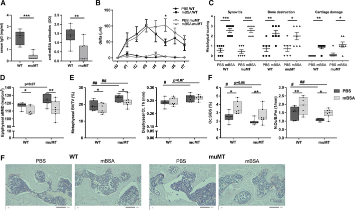
Fig. 6.

Mature B cells and immunoglobulins are dispensable for arthritis‐induced bone loss. Male muMT and WT littermate mice were immunized with mBSA. Antigen challenge was repeated intraarticularly after 7 days and knee joint swelling was measured daily over 7 days. (A) Serum IgG and anti‐mBSA antibodies were investigated at termination. Statistical analysis was performed using Student's t‐test, **p < 0.01, ***p < 0.001, n = 7. (B) Difference in swelling over the knee from baseline in micrometers (μm) in muMT and WT mice challenged with mBSA or PBS. Statistical analysis was performed at each measurement time point using Student's t test, *p < 0.05, n = 7. (C) Histological scoring (0–3) of synovitis, bone destruction, and articular cartilage damage 14 days after immunization. Statistical analysis was performed with Mann‐Whitney test in each genotype, *p < 0.05, **p < 0.01, ***p < 0.001, n = 7. (D) Epiphyseal aBMD was defined with DXA in the tibia. n = 6–7. (E) Diaphyseal Ct.Th and trabecular metaphyseal BV/TV in tibia were defined with high‐resolution μCT. (F) N.Oc/B.Pm and Oc.S/BS in the epiphyseal part of the tibia. n = 7. At termination Student's t test was performed between genotypes, WT and muMT, #p < 0.05, ##p < 0.01, and paired t test between intervention in the same mouse (non‐arthritic versus arthritic side), *p < 0.05, **p < 0.01. The interaction between the genotype (muMT and WT) and intervention (non‐arthritic and arthritic side) was calculated using a mixed‐model two‐way ANOVA. aBMD = areal bone mineral density; BV/TV = bone volume per total volume; Ct.Th = cortical thickness; mBSA = methylated bovine serum albumin; N.Oc/B.Pm = number of osteoclasts per bone perimeter; Oc.S/BS = osteoclast surface per bone surface; WT = wild‐type.
