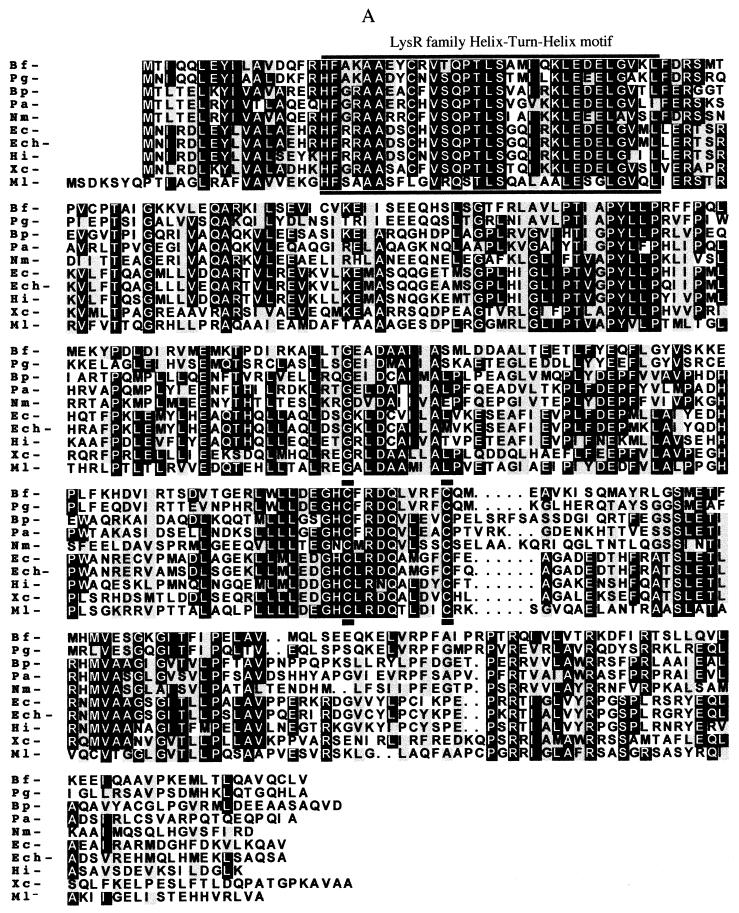
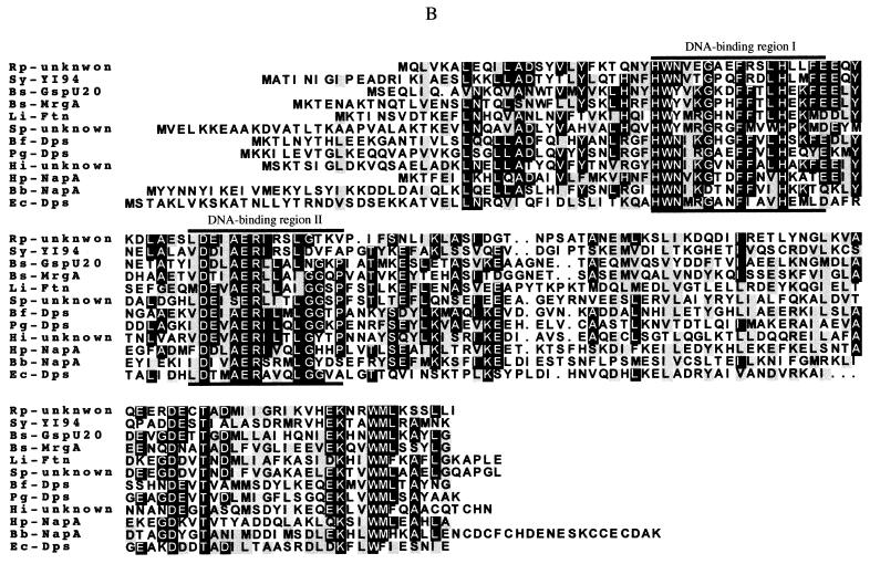
FIG. 1.
Multiple alignment of the B. fragilis (Bf) deduced amino acid sequences for OxyR (A) and Dps (B) with other bacterial OxyR and Dps homologue amino acid sequences, respectively. E. coli (Ec), B. burgdorferi (Bb), B. pertussis (Bp), B. subtilis (Bs), E. chrysanthemi (Ech), H. influenzae (Hi), H. pylori (Hp), L. innocua (Li), M. leprae (Ml), N. miningitis (Nm), P. gingivalis (Pg), P. aeruginosa (Pa), R. prowazekii (Rp), S. pneumoniae (Sp), Synechocystis sp. (Sy), and Xanthomonas campestris (Xc) sequences were used. Lines drawn above and below the amino acid sequences indicate the LyR helix-turn-helix DNA-binding nucleotide sequence motif and functional redox-active cysteine residues of E. coli OxyR C199 and C208 (29, 45) in panel A and the Dps protein signature consensus pattern associated with DNA binding properties (6, 43) in panel B. Consensus of at least 50% identical amino acid residues are labeled with black boxes. Conserved amino acid substitutions are depicted by grey boxes. The respective protein descriptions and GenBank accession numbers for the sequences are listed in Materials and Methods.


