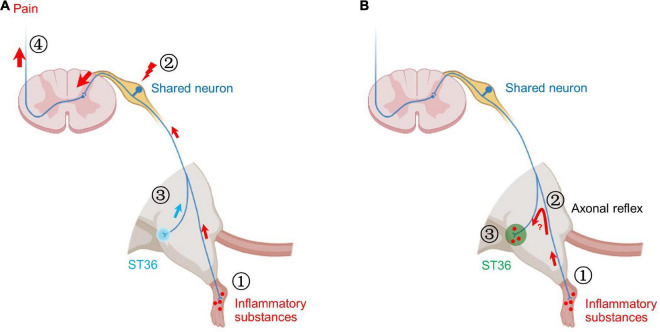
FIGURE 8.
Two possible mechanisms of shared neurons participating in acupoint sensitization. (A) ➀ The inflammatory substances induced by CFA in the hind paw plantar stimulate the terminals of shared neurons. ➁ The pain signals are continuously transmitted to the somata of shared DRG neurons, resulting in increased neuronal excitability. ➂ Mechanical stimulation of ST36 more readily activates shared neurons. ➃ More pain signals are transmitted to the brain. Behaviorally, the pain threshold of ST36 acupoint decreased. (B) ➀ The inflammatory substances induced by CFA in the hind paw plantar stimulate the terminals of shared neurons. ➁ By the axonal reflex mechanism, the nerve impulses are transmitted to ST36 acupoint via axon bifurcation. ➂ The persistent nerve impulses promote the release of pro-inflammatory molecules from the nerve terminal and recruit mast cells to aggregate, degranulate, etc. Locally enhanced neuroimmune responses lead to lower pain thresholds and enlarged receptive field of ST36 acupoint.

