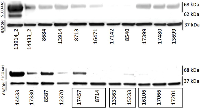
FIGURE 2.
Protein levels of OATP4A1 determined via Western blotting. The first two samples (13914_2 and 14433_2) are primary cell cultures containing mostly mesothelial cells. The three clusters on the bottom right (indicated with black rectangles) are cell lines derived from the same patient; all others are single cell lines generated from different patients. GAPDH was used as loading control.

