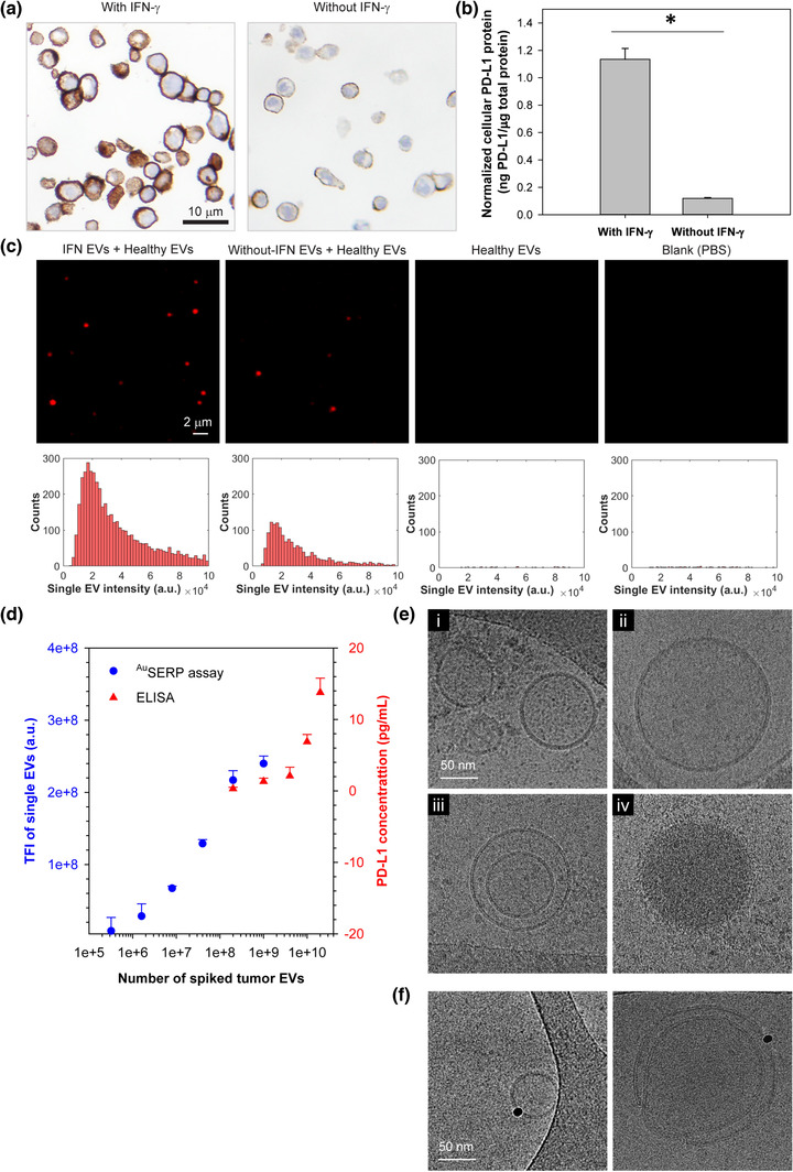
FIGURE 2.

In vitro model and characterization of cellular and single‐EV PD‐L1 protein. (a) Immunohistochemistry (IHC) of PD‐L1 protein in H1568 cells with/without interferon‐gamma (IFN‐γ) stimulation. Cell nuclei and PD‐L1 protein were stained blue (by haematoxylin) and brown (by Dako PD‐L1 IHC 22C3 pharmDx), respectively. (b) Quantification of PD‐L1 protein levels in H1568 cells measured by ELISA and normalized by the total protein expressed by the cells measured with a BCA Protein Assay. The data were expressed as mean ± SD; n = 3; *P < 0.001, Student's t‐test. (c) Representative TIRF microscopy images and their corresponding histograms of PD‐L1 protein expression on the surface of single EVs derived from H1568 cells with/without IFN‐γ stimulation in comparison to healthy donor EVs and PBS as controls. The single‐EV PD‐L1 protein signals were characterized with AuSERP using anti‐PD‐L1 antibodies and the TSA method. H1568 EVs were spiked in healthy donor EVs at a 1:1 ratio with 5 × 10¹⁰ particles/ml each. Healthy donor EVs were purified from healthy donor serum and then diluted in PBS to reach the target concentration. The images were cropped and enlarged from their original images, which are provided in Figure S3(b). (d) A performance evaluation of the AuSERP for PD‐L1 protein detection in comparison to ELISA (the average values are included in Table S3). EVs derived from IFN‐γ‐stimulated H1568 cells were spiked in healthy donor EVs at different concentrations ranging from 0 to 5 × 10¹⁰ particles/ml. The healthy donor EV concentration was kept constant at 5 × 10¹⁰ EVs/ml for all samples. The limit of detection (LOD) of AuSERP for PD‐L1 protein was ∼ 106 spiked tumour EVs, ∼ 1000 times lower than ELISA. The data were expressed as mean ± SD; n = 3. TFI, total fluorescence intensity; a.u., arbitrary units. (e) Cryogenic transmission electron microscopy (cryo‐TEM) images of EVs produced by IFN‐γ‐stimulated H1568 cells. (f) Cryo‐TEM images of immunogold labelled PD‐L1 protein on the EV surface
