
Figure 7. PCH mutations in CLP1 and the TSEN complex.
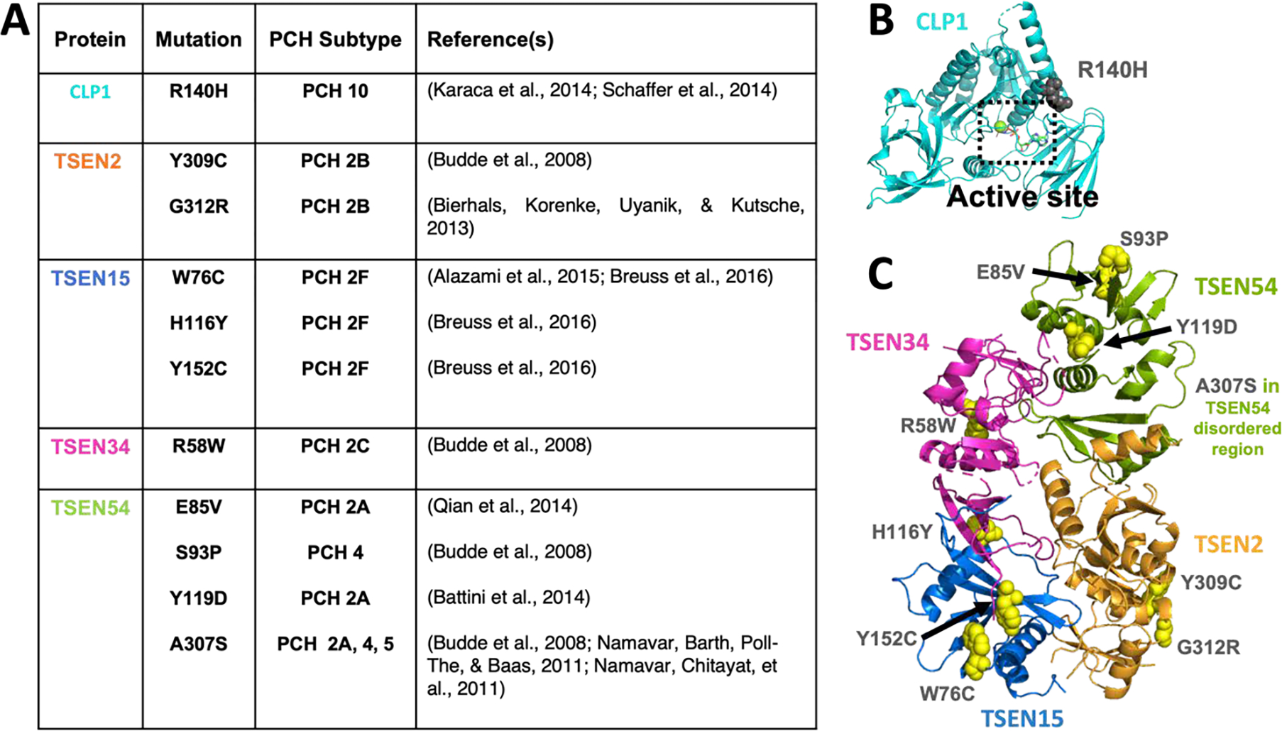
A) A table of PCH mutations in CLP1 and the TSEN proteins that have been characterized in the literature. B) CLP1 structure (PDB:4OHV) with the R140H PCH mutant in grey spheres and the active site boxed. C) The Alphafold TSEN complex model with disordered regions removed and with PCH mutations shown as yellow spheres and labelled. TSEN54’s mutation A307S was not in the regions shown.