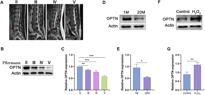
FIGURE 1.
OPTN level decreased in degenerated NP tissue and increased in H2O2-teated rat NPCs. (A) different degrees of IVDD MRI images of patients. (B–C) Western blot and quantification of OPTN in human NP tissue. (D–G) Western blot and quantification of OPTN in rat NP tissues and H2O2-teated NPCs. Experiments involving human NP specimen were performed as means ± SD of 5 times in duplicates. Experiments involving rat NP specimen were performed as means ± SD of 3 times in duplicates. *p < 0.05, **p < 0.01, ***p < 0.001, ns p > 0.05.

