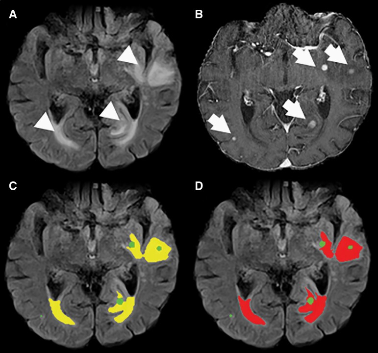
Figure 3.
Example of an MRI study with axial FLAIR and T1-weighted post-contrast images of a 71-year-old male patient with malignant melanoma and multiple BM in the institutional dataset (B, arrows and green) and perifocal edema (A, arrowhead). Our HD-BM algorithm detects the perifocal edema accurately (C, yellow) compared to the ground-truth segmentation (D, red).

