
Figure 3.
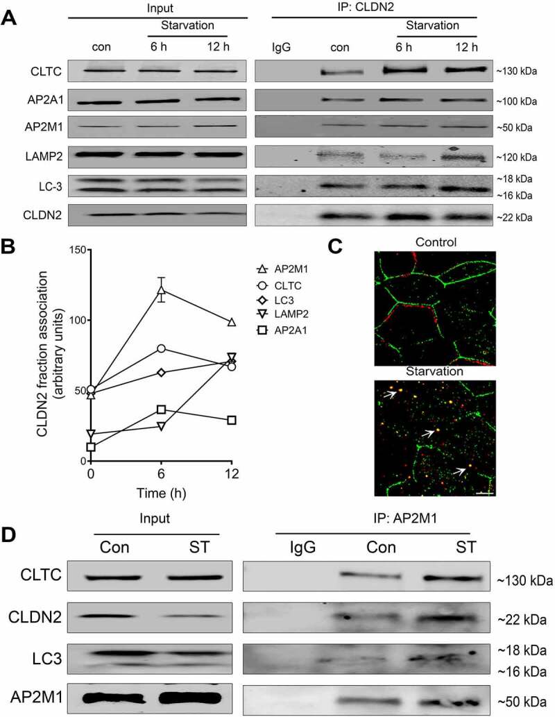
Interaction of CLDN2 with clathrin and autophagy apparatus. (A) Co-immunoprecipitation studies showed an increased association of CLDN2 with AP2M1, AP2A1, clathrin, LC3 during early starvation and lysosomal marker protein LAMP2 at the later 12-h time point. The negative control includes immunoprecipitation with control IgG. (B) Quantification of CLDN2 fraction associated with various clathrin and autophagy proteins, as shown in panel A. (C) Confocal immunofluorescence examination showed that, CLDN2 (green) migrated away from the cell membrane and increased cytoplasmic colocalization with clathrin (red) after starvation (yellow). White bar: 5 µm. (D) AP2M1 immunoprecipitates showed increased presence of CLDN2, LC3 and clathrin after starvation. Representation of ≥ 3 independent experiments.