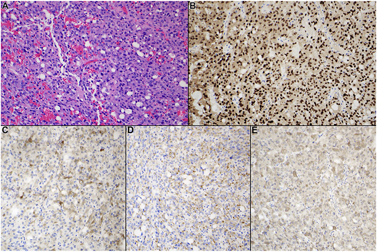
Figure 2.
Typical histological and immunohistochemical features of PIT-1–positive plurihormonal adenoma. All tumors included in this category demonstrated monomorphous features with no evidence of the acinar pattern of the normal pituitary gland. (A) Hematoxylin and eosin staining, (B) PIT-1 staining, (C) growth hormone staining, (D) prolactin staining, and (E) β-subunit of thyroid-stimulating hormone staining (original magnification ×200).

