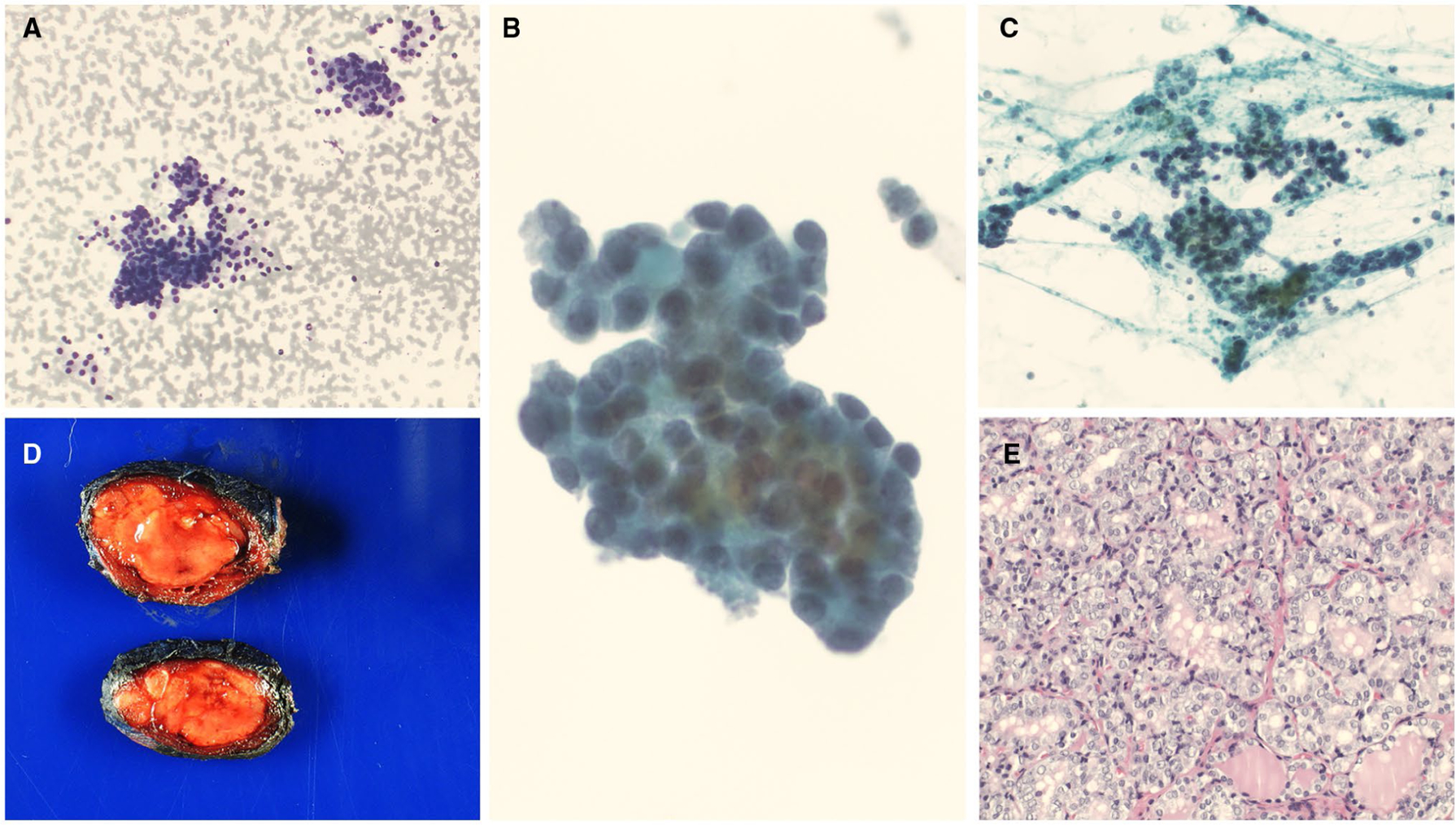
Figure 1.

HRAS mutations along with other molecular alterations. (A-C) A cytology evaluation showed clusters and groups of follicular cells in a microfollicular architecture. Some of the follicular cells showed nuclear crowding and slight nuclear enlargement (cytology diagnosis of follicular lesion of undetermined significance with nuclear atypia) ([A] Diff-Quik, ×100; [B] ThinPrep, ×400; [C] Papanicolaou stain, ×200). (D) A macroscopic examination showed a pale tan, somewhat circumscribed nodule (gross image). (E) A histologic evaluation showed papillary thyroid carcinoma, follicular variant (H & E, ×200).
