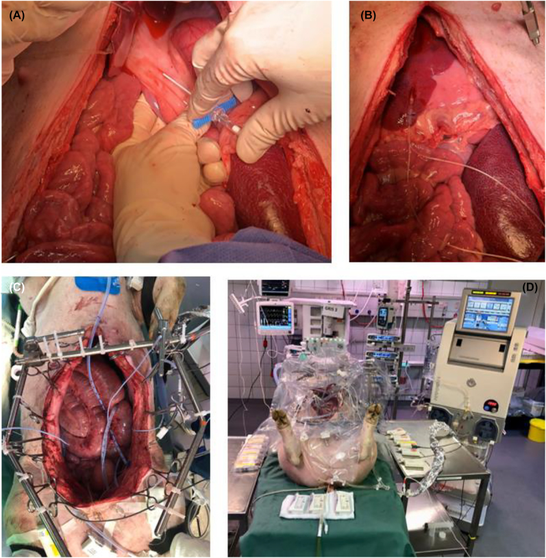
Figure 2:
Illustrative example of placement of microdialysis catheter.
(A) Illustrative example of placement of microdialysis catheter in hepatoduodenal ligament inserting an introducer. (B) Illustrative example of placement of a microdialysis catheter in the liver. The catheter is fastened to the liver tissue with a loose single suture to avoid displacement during sampling period. The same was done for all catheters. (C) Setup before HIPEC procedure seen from above. Placement of HIPEC tubes, HIPEC temperature probes and microdialysis catheters are shown. (D) Final setup placement of microdialysis pumps on tables, HIPEC performer system, and abdominal protection cover.

