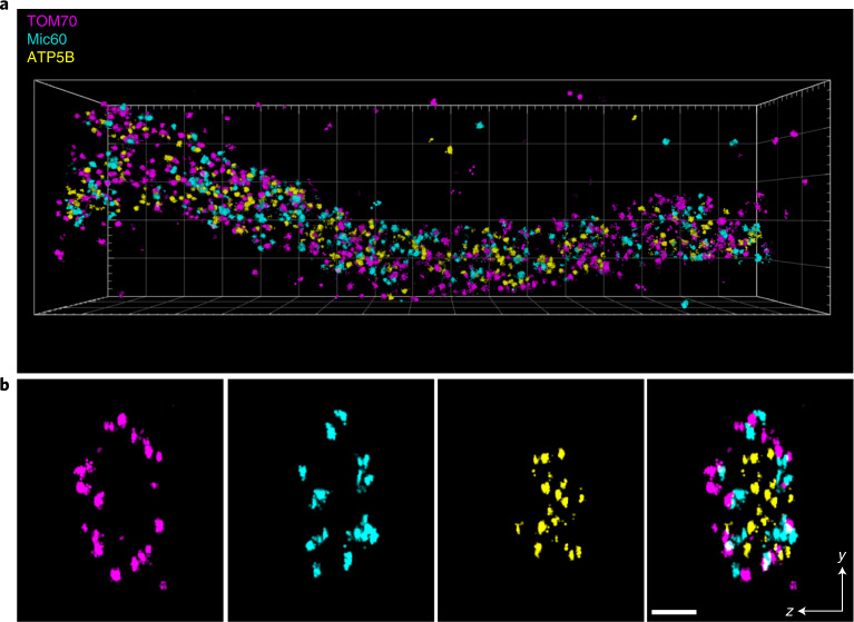
Fig. 2. 3D DNA-PAINT MINFLUX multiplexing.
U2OS TOM70-Dreiklang cells were fixed and immuno-labeled with an anti-GFP nanobody and anti-Mic60 and anti-ATP5B synthase antibodies. MINFLUX recordings of the three proteins were performed sequentially by adding and washing out the respective imager strands. Localizations of TOM70, Mic60 and ATP5B are displayed in magenta, cyan and yellow, respectively. a, View on a mitochondrial tubule. Size of the bounding box was 3.4 × 1 × 0.6 µm3. b, Cross section of the tubule shown in a. Thickness of the section 100 nm. Scale bar 100 nm.

