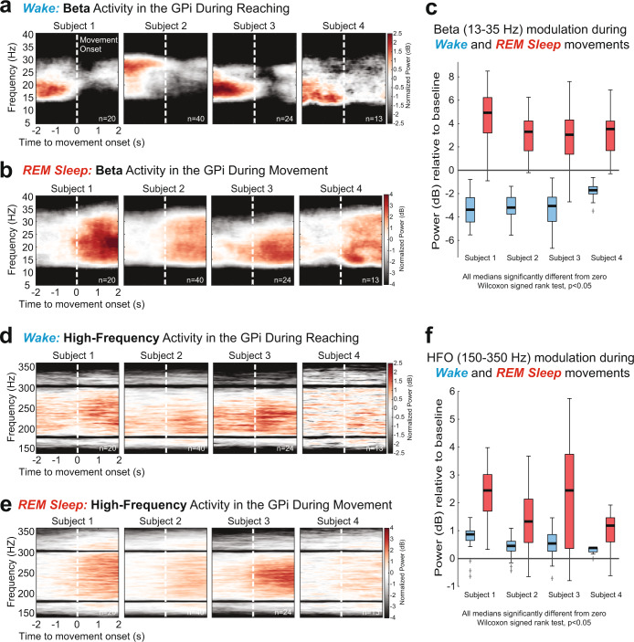
Fig. 1. Movement-related beta and high-frequency oscillations recorded from DBS leads in the GPi of PD patients in awake and REM sleep states.
a Trial-averaged spectrograms aligned to movement onset showing beta (13–35 Hz) desynchronization in the GPi during wakeful volitional movement (reaching task, see Methods). b Spectrograms showing beta synchronization in the GPi during REM sleep movements. c Distributions of beta band power modulation (relative to pre movement baseline) during wake movements (blue) and REM sleep movements (red). All data distributions were significantly different from zero (Wilcoxon signed rank (WSR) test, p < 0.05). d, e Trial-averaged spectrograms aligned to movement onset show synchronization of high-frequency oscillations (HFO, 150–350 Hz) in the GPi during wakeful and REM sleep movements, respectively. f Distributions of HFO band power modulation (relative to pre movement baseline) during wake movements (blue) and REM sleep movements (red). All data distributions were significantly different from zero (WSR test, p < 0.05). Boxplot elements: center line, median; box limits, upper and lower quartiles; whiskers, 1.5 × interquartile range; +sign, outliers.

