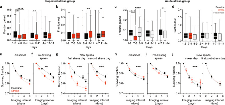
Fig. 4. Repeated and acute stress exposures have different effects on gain, loss and survival of dendritic spines in dCA1 PNs.
a Repeated stress exposure decreased the fraction of spines gained in the 1–4-day interval after stress onset (p7–8 = 0.0001, p8–9 < 0.0001, p9–11 = 0.0022, p11–14 = 0.7466, n = 124). b Repeated stress exposure increased the fraction of spines lost in the 4-to-7-day interval after stress onset (p7–8 = 0.7144, p8–9 > 0.9999, p9–11 = 0.0024, p11–14 = 0.0166, n = 124). c Acute stress decreased the fraction of spines gained immediately after stress exposure (p7–8 < 0.0001, p8–9 > 0.9999, p9–11 = 0.3483, p11–14 = 0.0182, n = 88). d Acute stress had no effect on spine loss (f, p7–8 > 0.9999, p8–9 = 0.3263, p9–11 = 0.8860, p11–14 = 0.2665, n = 88). Wilcoxon matched pairs signed ranks tests to 1–2, 2–4 and 4–7 respectively, p-values adjusted after Dunn’s correction for multiple comparisons Box plots: medians and quartiles of the distributions of fractional spine gain a, c or loss b, d per dendrite. e–g Repeated stress exposure decreased the survival of all e, pre-existing f and new g spines detected on the first (left) and second (right) days of stress. pAll = 0.003, pPre-existing = 0.001, pNewFirstday < 0.001, pNewSecondday < 0.001, nAll = 372, nPre-existing = 372, nNewFirstday = 312, nNewSecondday = 130. h–j A single exposure to stress increased the survival of all spines (h), but it did not affect pre-existing spines (i). Increase in spine survival was due to increased survival rate of new spines detected on the day of stress (j, left) but not on the following day (j, right). pAll < 0.001, pPre-existing = 0.251, pNewFirstday < 0.001, pNewSecondday = 0.861, nAll = 264, nPre-existing = 264, nNewFirstday = 177, nNewSecondday = 154. Mann–Whitney U-test between all non-1 Baseline versus Stress points. Circles: mean surviving fractions per dendrite. Error bars: s.e.m. Curves: single exponential decays fit to the data.

