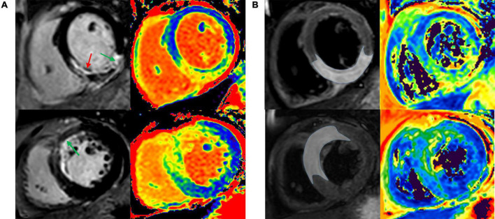
FIGURE 1.
Infarct region and AAR were validated by comparing T1 and T2 mapping against LGE and T2w-STIR. Representative images of patient with acute anterior and anteroseptal STEMI (A), and patient with acute inferior and inferoseptal STEMI (B). AAR, area at risk; LGE, late gadolinium enhancement; T2w-STIR, T2-weighted short tau inversion recovery; STEMI, ST-segment elevation myocardial infarction.

