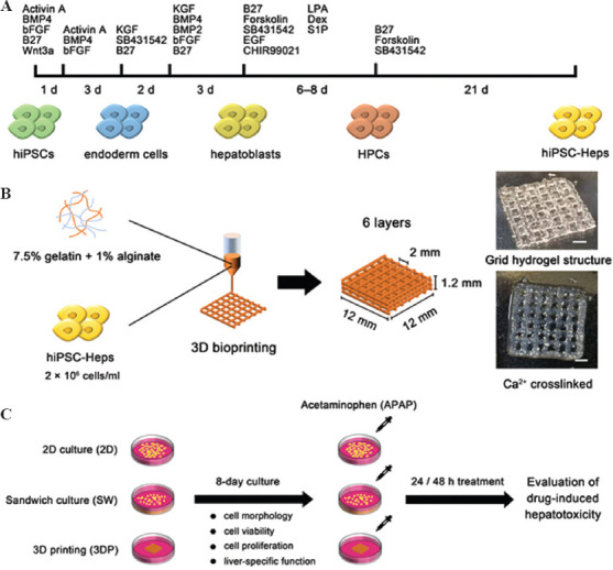
Figure 1.

Schematic figure of cell differentiation, bioprinting process, and experimental timeline. (A) Cell differentiation timeline from hiPSCs to hiPSC-Heps. (B) Bioprinting process: hiPSC-Heps were bioprinted using alginate-gelatin bioink to form a grid hydrogel structure as a hepatic tissue model. (C) Experimental timeline: The 2D-cultured (2D), the sandwich-cultured (SW), and the 3DP hepatic tissue model were cultured for 8 days, then treated with acetaminophen (APAP) for 24 or 48 h to evaluate drug-induced hepatotoxicity (Scale bar: 200 μm). hiPSCs, human-induced pluripotent stem cells; hiPSC-Heps, human-induced pluripotent stem cell-derived hepatocytes; HPC, hepatic progenitor cells.
