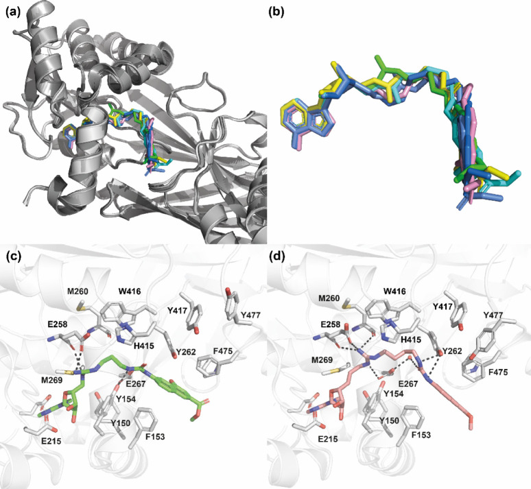
Figure 8.
Structures of mmPRMT4 in complex with compounds 12a–12c and 12f–12h (PDB IDs: 7PV6, 7PPY, 7PPQ, 7PU8, 7PUQ, and 7PUC, respectively). (a) Superimposition (done on protein backbones) of compounds (12a, 12b, 12c, 12f, 12g, and 12h) bound to subunit B of mmPRMT4. Each PRMT4 subunit is represented as a cartoon (shades of gray, lime, cyan, marine, yellow, gray, and pink ribbons), and compounds are represented as sticks (in lime, yellow, cyan, cornflower blue, sea blue, and pink, respectively). (b) Close-up view of bound compound conformations. (c) Binding interactions of compound 12a (lime sticks) with mmPRMT4 monomer B (ribbon). (d) Binding interactions of compound 12h (pink sticks) with mmPRMT4 monomer B (ribbon). Hydrogen bonds are shown as dashed lines. For clarity, N-terminal helices (residues 135–165) of PRMT4 are not shown.

