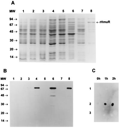
FIG. 7.
Expression, purification, and surface exposure of membrane-bound rHmuR. (A) Expression of rHmuR. The gene encoding the protein with the signal peptide was cloned into pCRT7/CT-TOPO and expressed in E. coli BL21(DE3)pLysE. Lanes are designated in the same manner as in Fig. 6A. (B) Identification of rHmuR. Whole-cell lysates and purified rHmuR were electrophoresed using SDS-PAGE and transferred onto a nitrocellulose membrane. Lanes are designated in the same manner as in Fig. 6B. The immunoblot was probed with anti-fusion protein antibody and detected using chemiluminescence staining. (C) Identification of rHmuR on the surface of E. coli BL21(DE3)pLyE cells (panel 1, E. coli harboring vector alone; panel 2, E. coli expressing membrane-bound rHmuR; panel 3, E. coli expressing rHmuR deposited in inclusion bodies). The dot blot was probed with antibodies against the fusion protein using cells before and 1 and 2 h after IPTG induction.

