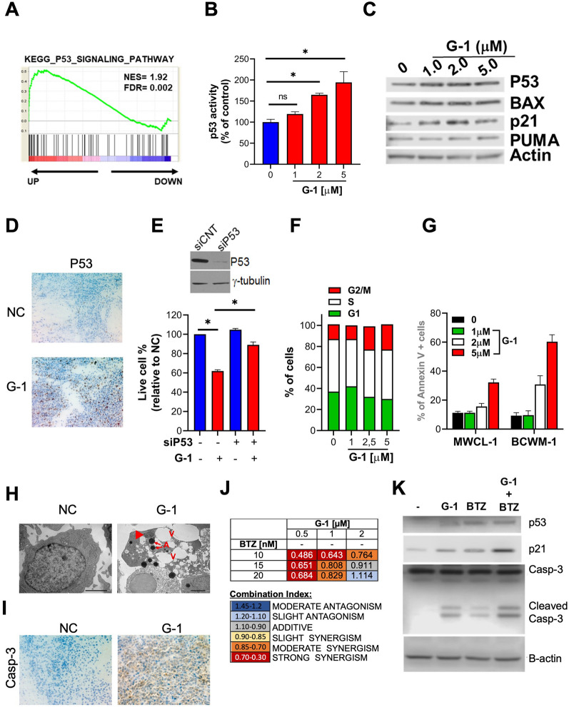
Fig. 2.
GPER1 pharmacological activation inhibits cell cycle progression and triggers apoptosis via inducing the p53 pathway. A GSEA performed 48 h after treatment with 1 μM G-1. B A p53 luminometric reporter assay was used to evaluate p53 transcriptional activity in G-1-treated BCWM-1 cells. C WB analysis of p53, p21, BAX, and PUMA in primary CD19+ WM cells treated with G-1 for 24 h. D Immunohistochemical staining for p53 (×20) in tumors sectioned on day 21 from vehicle- or G-1 [1 mg/kg] treated mice. Photographs are representative of one mouse receiving each treatment. E BCWM-1 cells were transfected with scrambled siRNAs (siCNT) or p53 targeting siRNAs and, after 24 h, were treated with vehicle or 1 μM G-1 for an additional 24 h and assessed for cell viability by CTG assay. WB analysis reports p53 knock-down in siP53-transfected cells. F FACS analysis of cell cycle phases of BCWM-1 cells 24 h after treatment with vehicle or G-1. G Annexin V staining of BCWM-1 cells 24 h after treatment with vehicle or G-1. H TEM analysis of BCWM-1 cells treated with G-1 (1 μM) or DMSO (NC). Control cells appear well-preserved with intact mitochondria, orderly chromatin folding and a clear nuclear membrane. Apoptotic cells become pyknotic with many electron-transparent vacuoles (V), chromatin (arrowhead) and cytoplasm condensation (increase in electron density of cytoplasmic matrix and organelles) and formation of apoptotic bodies (A). I Immunohistochemical staining for caspase 3 (×20) in tumors sectioned on day 21 from vehicle- or G-1 [1 mg/kg] treated mice. J Table showing combination indexes resulting from combinatorial treatments of BCWM-1 with G-1 and bortezomib (24 h time point). K WB analysis of p53, p21 and caspase 3 in BCWM-1 cells treated with [0.5 μM] G-1 and 10 nM bortezomib (BZ)

