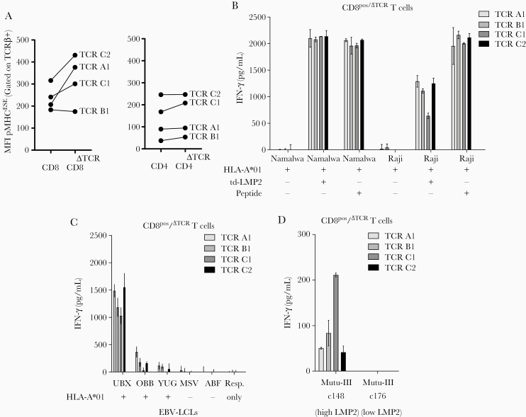
Figure 4.
CD8+/ΔTCR T cells transduced with Epstein-Barr virus Latent Membrane Protein 2 ESEERPPTPY (EBV-LMP2ESE)–specific T-cell receptors (TCRs) effectively recognize endogenously processed and presented LMP2ESE peptide. A, Mean fluorescence intensity (MFI) of peptide major histocompatibility complex (pMHC) EBV-LMP2ESE tetramer binding was assessed for CD8+ (left) and CD4+ (right) T cells with and without (ΔTCR) endogenous expressing TCRs. B, CD8+/ΔTCR T cells transduced with EBV-LMP2ESE–specific TCRs were stimulated for 16 hours with HLA-A*01:01–transduced EBV-associated malignant cell lines Namalwa and Raji in a responder-to-stimulator ratio of 1:5. These cell lines were additionally transduced with LMP2 and the cell lines without LMP2 were exogenously pulsed with 10–6 M of EBV-LMP2ESE peptide. C, CD8+/ΔTCR T cells transduced with EBV-LMP2ESE–specific TCRs were stimulated for 16 hours with HLA-A*01:01–positive and HLA-A*01:01–negative EBV-transformed lymphoblastoid cell lines (LCLs) that express LMP2 under physiological conditions in a responder-to-stimulator ratio of 1:5. D, CD8+/ΔTCR T cells transduced with EBV-LMP2ESE–specific TCRs were stimulated for 16 hours with 2 HLA-A*01:01–transduced Burkitt lymphoma mutu-III cell lines that were expected to express LMP2 under physiological conditions. However, mutu-III subclone c148 expressed LMP2 >100-fold higher compared to subclone c176. Data shown are from separate experiments carried out in triplicate (B, C, and D) with T cells from donor H.

