. 2022 Sep 13;11:e80813. doi: 10.7554/eLife.80813

Figure 2. Analysis of PfA-M17 localization and expression over the lifecycle.

Figure 2.

(A) Immunofluorescent analysis of PfA-M17-HAglmS parasites in the three distinct lifecycle stages fixed with 90:10 acetone:methanol and probed with anti-HA and DAPI. (B) Saponin-lysed mixed stage PfA-M17-HAglmS parasites were sequentially lysed in the buffers indicated from left to right and analyzed by Western blotting. Insoluble material represents the remaining pellet after lysis in 1% Triton X-100. GAPDH, HSP101, and EXP2 serve as controls for cytoplasmic, membrane-associated, and integral membrane proteins respectively. Blot is representative of three biological replicates. (C) Western blot analysis of endogenous PfA-M17 expression in Pf3D7 wildtype parasites over the erythrocytic cycle probed with anti-M17 antibodies. HSP101 serves as a loading control.

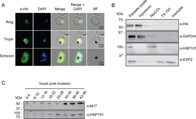
Figure 2—source data 1. Original immunoblot presented in Figure 2B (anti-HSP101 blot).
Figure 2—source data 2. Marked up original immunoblot presented in Figure 2B (anti-HSP101 blot).
Figure 2—source data 3. Original immunoblot presented in Figure 2B (anti-GAPDH blot).
Figure 2—source data 4. Marked up original immunoblot presented in Figure 2B (anti-GAPDH blot).
Figure 2—source data 5. Original immunoblot presented in Figure 2B (anti-HA blot).
Figure 2—source data 6. Marked up original immunoblot presented in Figure 2B (anti-HA blot).
Figure 2—source data 7. Original immunoblot presented in Figure 2B (anti-EXP2 blot).
Figure 2—source data 8. Marked up original immunoblot presented in Figure 2B (anti-EXP2 blot).
Figure 2—source data 9. Original immunoblot presented in Figure 2C.
Figure 2—source data 10. Marked up original immunoblot presented in Figure 2C.