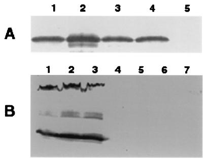
FIG. 2.
Immunoblot analysis of pilus- and fibril-specific proteins. All samples were separated by SDS–10% PAGE (28). The presence of pilus- and fibril-specific proteins was analyzed using anti-PilA serum (A) and monoclonal antibody MAb2105 (B) as primary antibodies, respectively. In panel A, samples from 5 × 10⁸ cells were loaded on each lane. Whole-cell lysate from 5 × 10⁷ cells was loaded on each lane in panel B. Panel A lanes: 1, SW505; 2, SW501; 3, SW504; 4, DK1622; 5, DK1253. Panel B lanes: 1, DK1622; 2, DK1253; 3, DK1300; 4, SW505; 5, SW501; 6, SW504; 7, DK3470.

