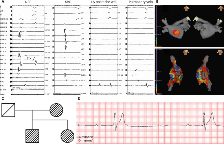
Figure 3. Electrophysiologic study and ECG. Intracardiac electrogram during sinus rhythm with HV interval 73 ms. In contrast to the absence of endocardial signals in LA posterior wall and pulmonary vein, SVC showed relatively preserved signals in intracardiac electrograms (A). The left atrial voltage map with final ablation site at RA (B). A genotype pedigree representing first-degree relatives (C). Follow-up Holter showing paroxysmal complete AV block in November 2021 (D).
AV = atrioventricular; aVF = augmented vector foot; HV = His-ventricular; LA = left atrial; NSR = normal sinus rhythm; RA = right atrium; SVC = superior vena cava.

