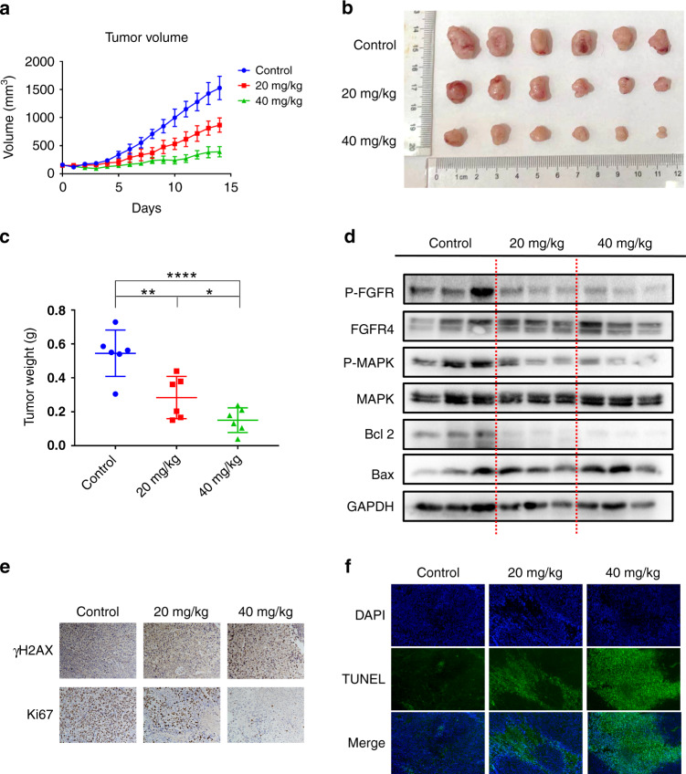
Fig. 7. The anti-tumour activity of F1-7 in vivo.
a Tumour volume was measured every day. b The graph of tumours in different groups. c The weight of the tumours was measured. d The tumour tissues were extracted in lysis buffer, and western blot analysis was performed. e Representative immunohistochemical staining images of cell proliferation marker (Ki-67) and DNA damage marker (γH2AX) in tumour tissues. f TUNEL staining results of the tumour tissues.

