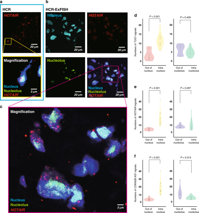
Fig. 5. The HCR-ExFISH reveals intracellular colocalization of lncRNAs.
a, b LncRNA HOTAIR signals by conventional HCR (a) and nanoresolution HCR-ExFISH (b). Images are acquired using confocal microscopy. c High-magnification image of the boxed region in (b). d–f Violin plot shows the spatial heterogeneity of lncRNA TUG1 (d), HOTAIR (e) and CDKN2B-AS1 (f) signals in subcellular localisation, compared using the two-tailed Student’s t test.

