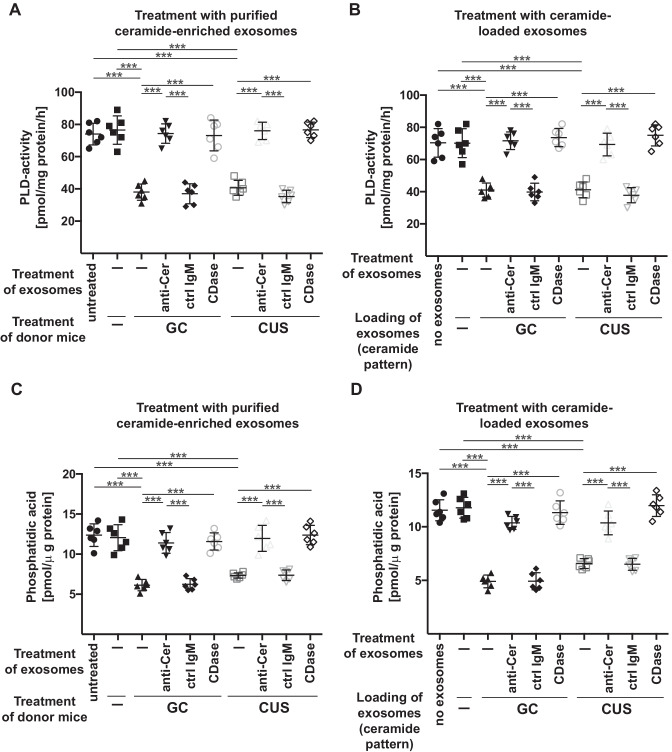
Fig. 6.
Incubation of endothelial cells with exosomes isolated from stressed mice or with ceramide-loaded exosomes inhibit PLD and reduce phosphatidic acid. A and C bEnd.3 cells were incubated with exosomes isolated from the blood plasma of wild-type mice, which were left untreated (-) or stressed with glucocorticosterone (GC) or chronic unpredictable stress (CUS). Purified exosomes were treated ex vivo with ceramide IgM antibodies clone S58-9 (anti-Cer), control immunoglobulin M (ctrl IgM), or recombinant ceramidase (CDase) or were left untreated (-), pelleted by ultracentifugation, resuspended in H/S and added to bEND3 endothelial cells. Control mice (Ctrl) were completely left untreated (-). Cells were incubated for 2 h and endothelial PLD activity A and endothelial phosphatidic acid concentrations C were determined. B and D Exosomes were purified from the blood plasma of untreated mice, loaded with C22, C24, and C24:1 ceramide in the amounts and ratio as these species are present in exosomes isolated from glucocorticosterone (GC)-treated mice or in exosomes isolated from mice treated with chronic unpredictable stress (CUS) and washed. bEnd3 endothelial cells were incubated with these exosomes for 2 h and PLD activity B and phosphatidic acid concentrations D in endothelial cells were determined. Shown are the mean ± SD from each 6 experiments/group. ***P < 0.001, ANOVA and post hoc Tukey test

