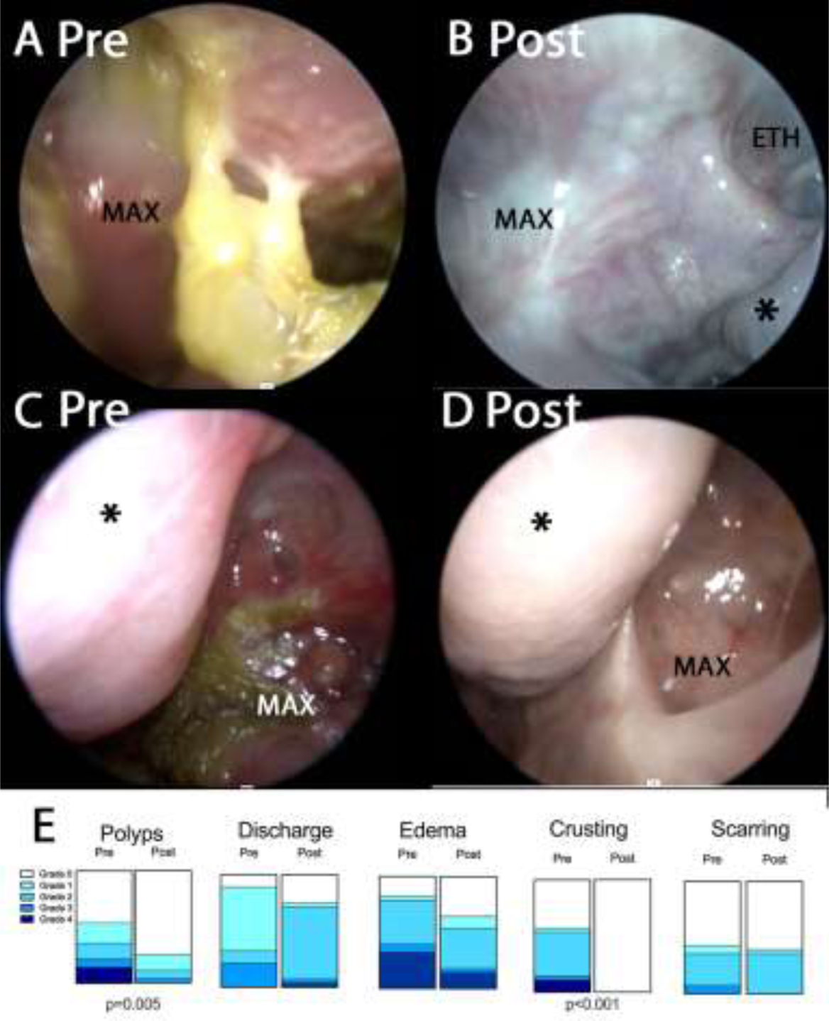
Figure 2:

Rigid sinonasal endoscopic photographs pre (A, C) and post (B, D) ELX/TEZ/IVA treatment. Fig 2A–B: Right maxillary-ethmoid. Note adherent yellow mucus present in A prior to treatment. Fig 2C–D: Left maxillary. Note erythema, mucosal edema and discharge in C prior to treatment. * indicates middle turbinate. MAX indicates maxillary sinus. ETH indicates ethmoid. Fig 2E. Lund-Kennedy items scored before and after treatment with ELX/TEZ/IVA. p-values shown from McNemar test comparing grade 0 to grades >0.
