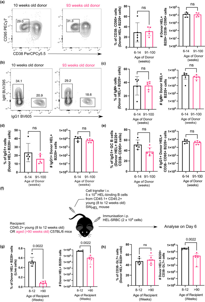
FIGURE 7.

B cells from aged donor mice do not have defects in participating in the GC 10 days after immunization in young recipient mice and fewer B cells from young donor mice are recovered when transferred into aged recipient mice. (a) Representative flow cytometric plots of CD38− CD95+ GC B cells gated on donor cells from 10‐week‐old or 93‐week‐old donor mice in recipient spleens on day 10 post‐transfer and immunisation. Numbers adjacent to gates indicate percentage of donor HEL+ B220+ cells. Percentage and number of donor‐derived GC B cells (Donor HEL+ B220+ CD38− CD95+) are plotted on the graphs on the right. (b) Representative flow cytometric plots of IgM+ and IgG1+ donor HEL+ B cells. Numbers adjacent to gates indicate percentage of donor HEL+ B220+ cells. (c, d) Percentage and number of (c) IgM+ and (d) IgG1+ B cells derived from donor cells from young or aged mice in recipient spleens 10 days post‐transfer and immunisation. (e) Graph showing the percentage and number of IgG1+ GC B cells out of total Donor HEL+ B220+ CD38− CD95+ cells. (f) Schematic diagram of adoptive transfer of B cells from young SWHEL mice into young (8–12 weeks old) or aged (>90 weeks old) mice in which B cells response was analysed 6 days post‐transfer and immunisation. (g) Percentage and number of donor HEL+ B220+ cells in spleens of young or aged recipient mice 6 days post‐transfer and immunisation. (h) Percentage and number of donor‐derived GC B cells (Donor HEL+ B220+ CD38− GL7+) in spleens of young or aged recipient mice 6 days post‐transfer and immunisation. Bar graphs show the results of one of two independent experiments (n = 5–6 per group/experiment). Bar height corresponds to the mean, error bars indicate standard deviation and each symbol represents one biological replicate. Statistics were calculated using the unpaired Mann–Whitney U test.
