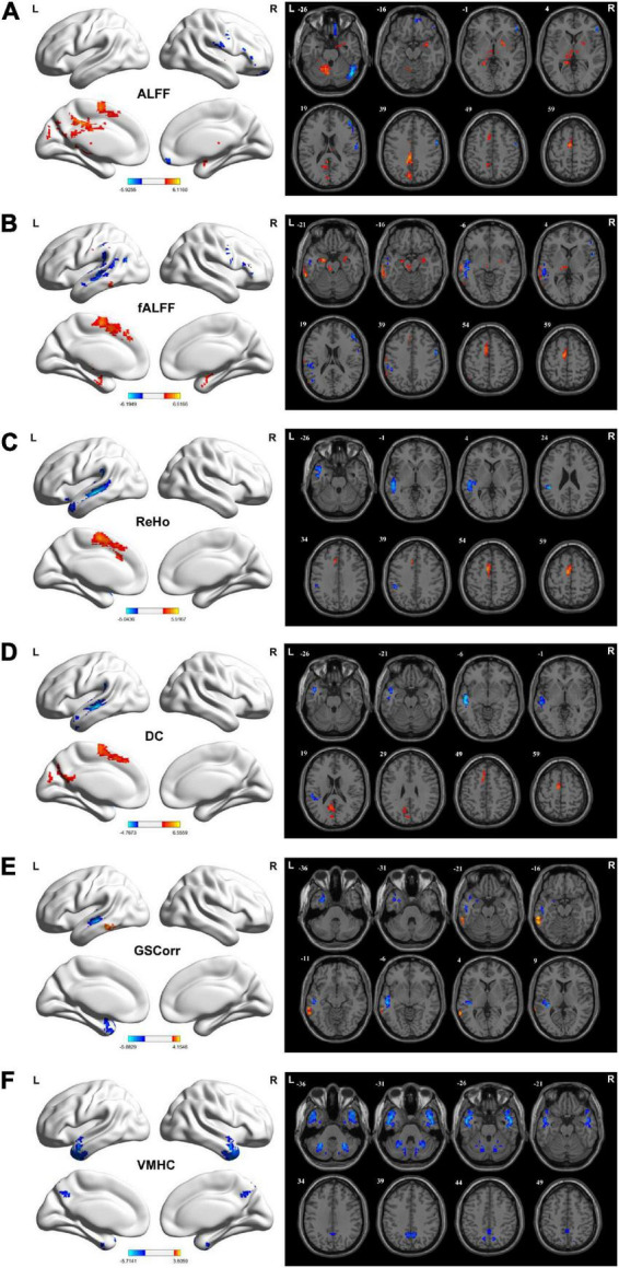
FIGURE 1.

Brain regions showing considerably altered static ALFF (A), fALFF (B), ReHo (C), DC (D), GSCorr (E), and VMHC (F) between TLE and HCs groups. GRF corrected; voxel-wise P < 0.005, cluster-wise P < 0.05. The color bar indicates the t-value. Warm colors indicated increased indicator values, while cold colors indicated decreased indicator values. ALFF, amplitude of low-frequency fluctuation; DC, degree centrality; fALFF, fractional ALFF; GRF, Gaussian random field theory; GSCorr, global signal correlation; ReHo, regional homogeneity; TLE, temporal lobe epilepsy; VMHC, voxel-mirrored homotopic connectivity.
