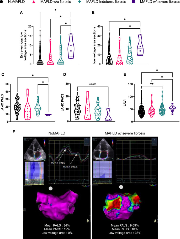
Figure 1.
Left atrial structural and electrical remodeling parameters. Number of low-voltage area extra-venous (A) or total (B); peak atrial longitudinal strain (C) and peak atrial contraction strain (D); left atrial volume indexed to body surface area (E). Representative echography, strain values, and bipolar voltage maps (low-voltage cutoff: 0.5 mV) (F). NoMAFLD: FLI < 60; MAFLD w/o fibrosis: FLI > 60 and NFS < 1.455; MAFLD ind. fibrosis: FLI > 60 and −1.455 < NFS < 0.675; MAFLD w/severe fibrosis: FLI > 60 and NFS > 0.675. Kruskal–Wallis test followed by Dunn’s post-hoc test. *p < 0.05; **p < 0.01. MAFLD, metabolic dysfunction-associated fatty liver disease; LA, left atria; PALS, peak atrial longitudinal strain; PACS, peak atrial contraction strain; LAVI, left atrial volume index.

