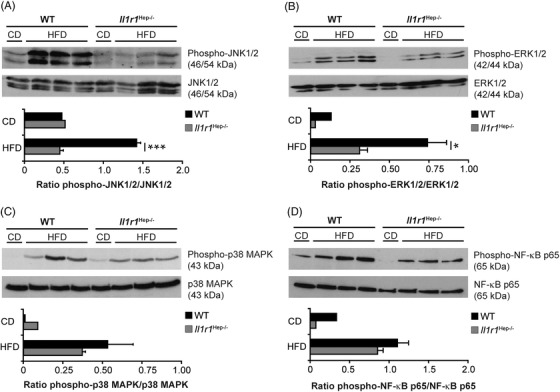
FIGURE 5.

HFD‐induced alterations of MAPK and NF‐κB p65 signalling in the liver of Il1r1 Hep−/– and WT mice. Immunoblotting of (A) phospho‐JNK1/2 (Thr183/Tyr185), (B) phospho‐ERK1/2 (Thr202/Tyr204), (C) phosho‐p38 MAPK (Thr180/Tyr182), (D) phospho‐NF‐κB p65 (Ser536) and total (A) JNK1/2, (B) ERK1/2, (C) p38 MAPK, and (D) NF‐κB p65 protein in liver whole tissue lysates of the different experimental groups after 12 weeks of dietary feeding. In A‐D representative immunoblots with densitometric analysis are shown. *p < .05, ***p < .001 for Il1r1 Hep−/– versus WT using unpaired, two‐tailed Student's t‐test (A and B). There was no statistically significant difference between Il1r1 Hep−/– and WT mice on HFD with respect of the parameters in C and D
