FIGURE 6.

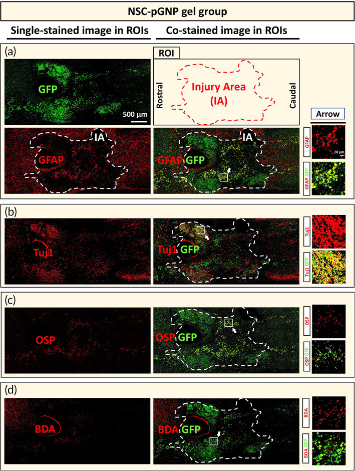
Cellular differentiation of grafted neural stem cells (NSCs) under the spinal cord injury condition for the NSC‐positively charged gold nanoparticles gel group. Horizontally sectioned specimens were stained with the green fluorescent protein (GFP) antibody. Each GFP‐stained section was co‐stained with glial fibrillary acidic protein (GFAP) (36th and 40th), Tuj1 (37th and 41st), oligodendrocyte specific protein (OSP) (38th and 42nd), or biotinylated dextran amines (BDA, 39th and 43rd). Representative tile scan images (also designated as regions of interest [ROIs], 4500 × 2000 μm2) of samples co‐labeled with (a) GFAP/GFP, (b) Tuj1/GFP, (c) OSP/GFP, and (d) BDA/GFP are shown. Scale bar of the ROI image is 500 μm (IA = Injury Area). Arrows indicate randomly designated regions for higher magnification views. The designated region is 170 × 170 μm2 (Scale bar: 20 μm)
