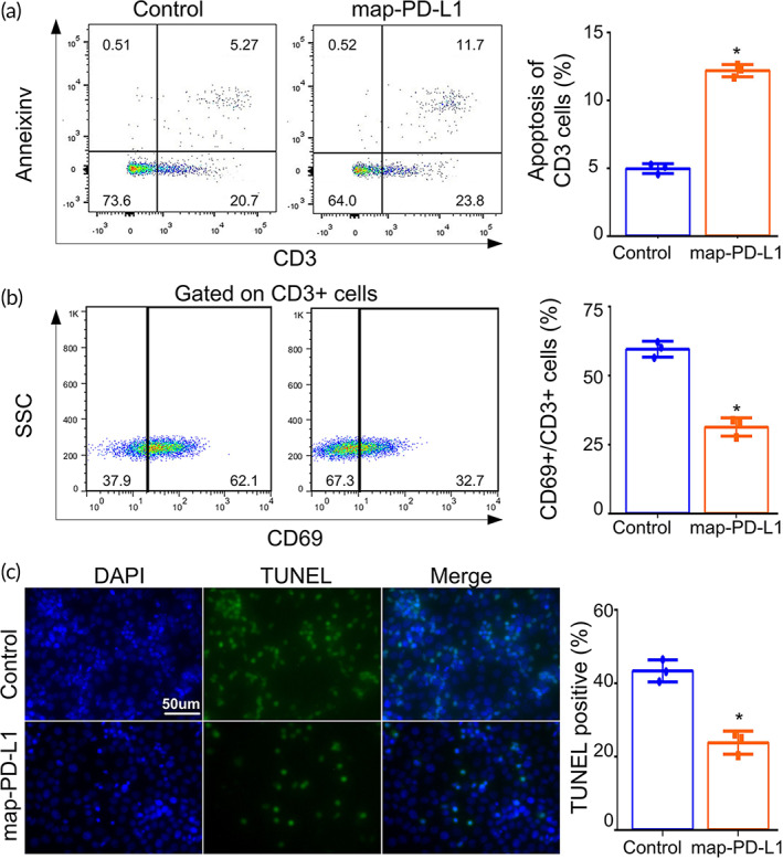
FIGURE 4.

In vitro experiments to verify the biological functions of map‐PD‐L1. Spleen lymphocytes were cocultured with rgEC. For the T cell activation assay, cells were cocultured with anti‐CD3 and anti‐CD28 antibodies for 1 day as a stimulation protocol. In the experimental group, map‐PD‐L1 was anchored to rgEC. Apoptosis (a) and activation (b) of T cells were analyzed by PI and annexin V staining, and CD69 molecular markers, respectively. (c) Detection of rgEC apoptosis after coculture with spleen lymphocytes using a TUNEL Apoptosis Assay Kit. Original magnification, ×400. map‐PD‐L1, membrane‐anchored‐protein PD‐L1; rgEC, rat glomerular endothelial cell
