FIGURE 12.

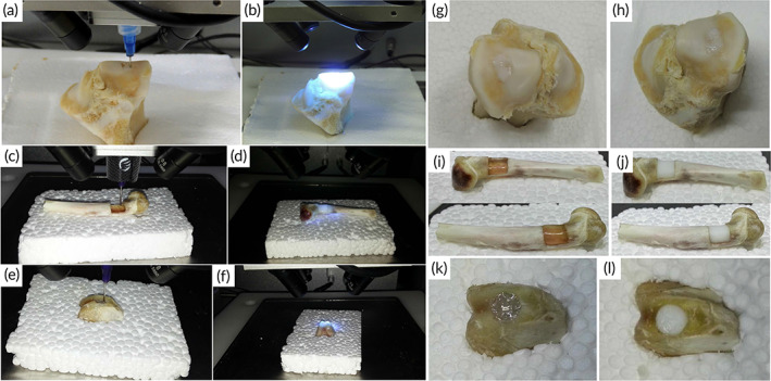
On chondral, bone, and osteochondral abnormalities, a 3D bioprinting and photopolymerization process was used. (a) In situ 3D bioprinting using m‐HA hydrogel to repair a chondral lesion. (b) UV light exposure of printed m‐HA hydrogel (c) in situ 3D bioprinting with alginate hydrogel to repair a bone defect. (d) UV light exposure of printed alginate hydrogel. (e) In situ 3D bioprinting with alginate hydrogel to repair an osteochondral lesion. (f) UV light exposure of printed alginate hydrogel. (g,h) Before and after photopolymerization, the color of the m‐HA hydrogel used to repair the chondral defect was milky white. (i) Before photopolymerization, the alginate hydrogel used to repair the bone defect was transparent. (j) After a few seconds of exposure to UV radiation, the hue of the alginate hydrogel became milky white. (k) Before photopolymerization, the alginate hydrogel used to repair the osteochondral lesion was transparent. (l) After a few seconds of exposure to UV light, the hue of the alginate hydrogel became milky white. The chondral, bone, and osteochondral deficiencies were all completely repaired. Source: All figures were reprinted with permission from reference 85
