Figure 1.
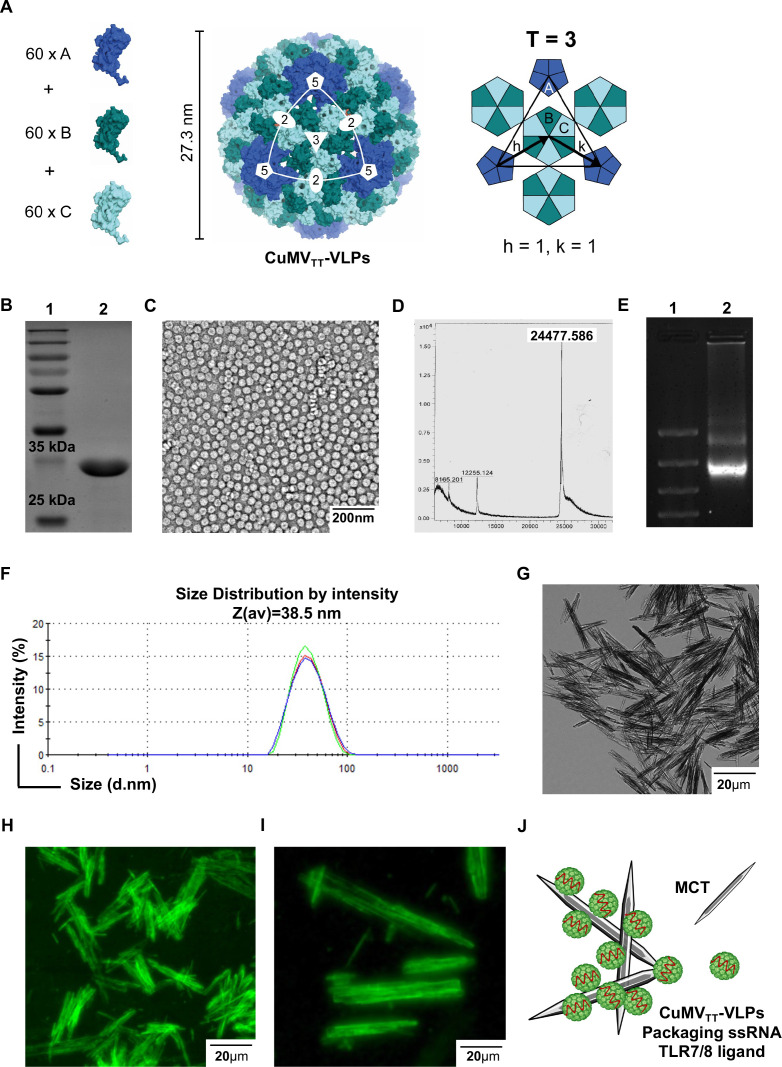
CuMVTT nanoparticles decorate the surface of MCT micron-sized crystals. (A) Geometry of the icosahedral CuMVTT VLPs illustrating the capsid protein in T=3 configuration. VLPs are formed of 60 of each subunits A, B and C (illustrated here in shades for clarity). (B) SDS-PAGE analysis of CuMVTT VLPs, lane 1: protein size marker (Thermo Scientific), lane 2: 6 µg VLPs. A monomer of CuMV-protein has a size of ~(25–30 kDa). (C) Electron microscopy image of purified VLPs, VLP sample (1.5 mg/mL) examined by JEM-100C electron microscopy, VLP’s size between 26 and 28 nm. (D) Mass spectrometry analysis confirmed a major peak of ~24,477.586. (E) Agarose gel analysis of the VLPs, lane 1: DNA marker (Thermo Fisher), lane 2: 10 µg purified VLPs, VLP sample (1.5 mg/mL). (F) Dynamic light scattering analysis of the VLP nanoparticles, VLP sample (1.5 mg/mL). Three measurements were performed and analyzed by DTS software. (G) MCT crystals visualized with light microscopy, exhibiting a micron size of ca=4.5 µm in length. (H) CuMVTT VLPs labeled with AF488 and formulated with MCT micron-sized adjuvant, referred to as immune-enhancer, ×40 objective scale bar 20 µm. (I) CuMVTT VLPs labeled with AF488 and formulated with MCT micron-sized adjuvant, ×40 objective scale bar 20 µm. (J) A cartoon illustrating CuMVTT VLPs; packaging ssRNA (in red) and decorating MCT crystals. CuMVTT, cucumber mosaic virus-like particles incorporating a tetanus toxin peptide; MCT, microcrystalline tyrosine; VLP, virus-like particle.

