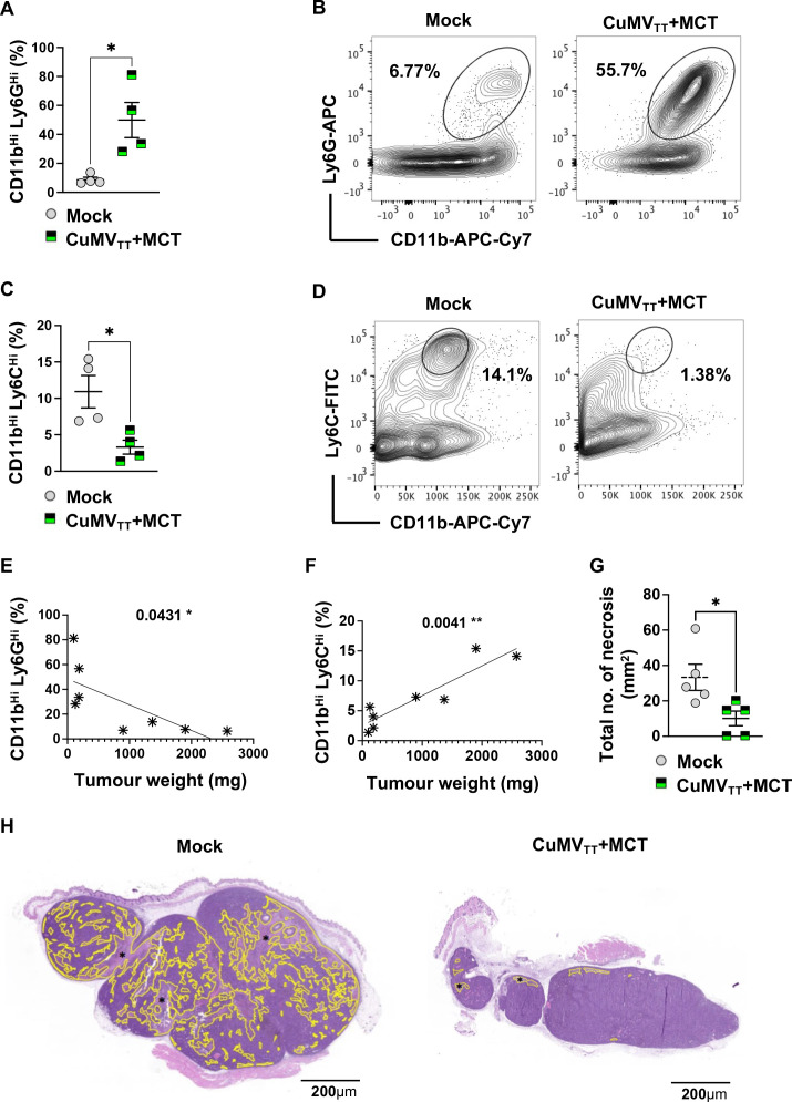
Figure 4.
Intratumoral administration of CuMVTT+MCT forms a depot, enhances inflammation and reduces tumor necrosis. (A) Percentage (%) of CD11bHi Ly6GHi cells in tumors; each dot represents an individual tumor. (B) Representative FACS plots showing the percentage (%) of CD11bHi Ly6GHi cells in a tumor. (C) Percentage (%) of CD11bHi Ly6CHi cells in tumors; each dot represents an individual tumor. (D) Representative FACS plots showing the percentage (%) of CD11bHi Ly6CHi cells in a tumor. (E) Correlation between percentage (%) of CD11bHi Ly6GHi cells and tumor weight (in mg). (F) Correlation between percentage (%) of CD11bHi Ly6CHi cells and tumor weight (in mg). (G) Total number of necrosis (mm2); each dot represents an individual tumor. (H) Example of a mock tumor (left): necrosis comprises 30% of the tumor section, example of a treated tumor with the immune-enhancer (right): necrosis comprises 2% of the tumor section. Necrosis is indicated in yellow (*). The entire tumor cross section was assessed for necrosis, H&E staining. Statistical analysis (A, C, G) by Student’s t-test (mean±SEM). Statistical analysis (E, F) by linear regression. n=4 (A–C) and n=5 (E, F, G), one representative of two similar experiments is shown. MCT, microcrystalline tyrosine.

