Figure 1.
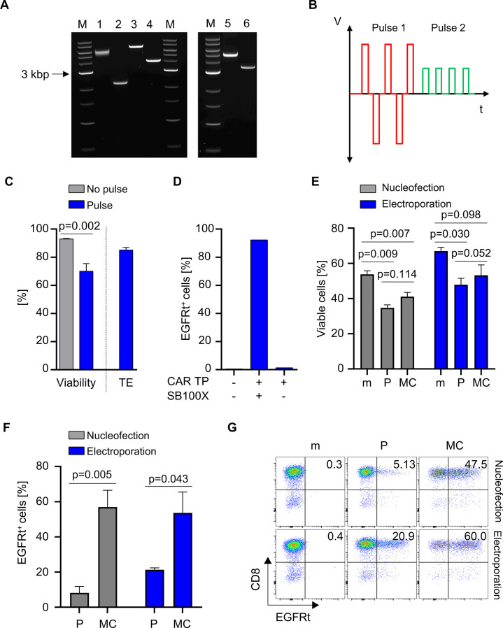
Small scale transfection of T cells using bi-pulse electroporation. (A) Restriction digestion analysis of conventional plasmids and MCS. Lane 1: SB100X plasmid digested with Sac I; Lane 2: SB100X MC digested with Pac I; Lane 3: PT2 CD19 CAR plasmid digested with Nhe I; Lane 4: PT2 CD19 CAR MC digested with Pac I, Lane 5: PT2 EGFP plasmid digested with Nhe I; Lane 6: PT2 EGFP MC digested with Pme I; lanes M: 1 kb DNA ladder (NE). (B) Schematic representation of applied bi-pulse system (red=first pulse, green=second pulse). (C) Viability and transfection efficiency (TE) of GFP-MC electroporated (Pulse) or non-electroporated (No pulse) T cells using the established bi-pulse system. (D) Episomal CAR-expression from MC vectors. T cells were electroporated with CD19 CAR transposon donor MC vector (CAR TP) in the presence or absence of transposase (SB100X), CAR-expression was assessed on day 6. (E–G) CD8 T cells were (co-)electroporated with mock (m, no DNA) or CD19 CAR and SB100X either encoded on plasmid (P) or MC in equimolar amounts using the two different electroporation systems. (E) viability, (F) transfection efficiency and (G) a representative flow cytometry analysis of anti-EGFRt staining 12 days after electroporation are shown. (C), (E, F) show mean+SD from N=3 donors. Statistical analysis was performed using paired t-test.

