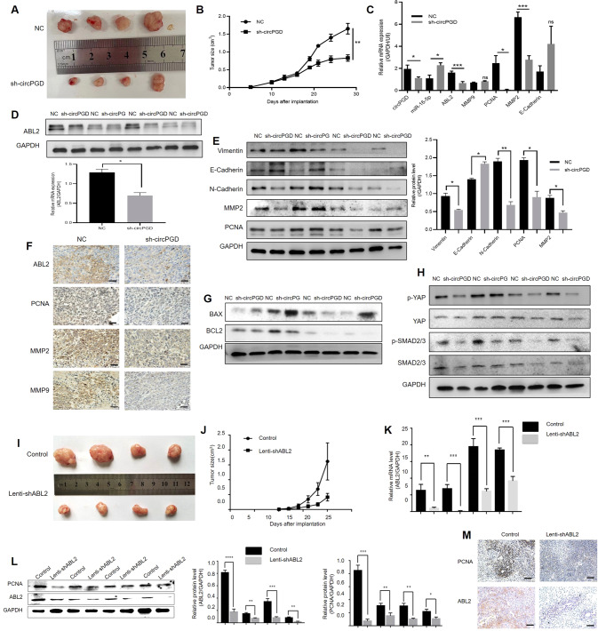
Fig. 7. CircPGD and ABL2 promotes GC cell growth in vivo.
A and I Images showing MGC-803 cells induced subcutaneous xenografts. B and J MGC-803 cells induced tumor volumes measured every 3 days for 6–8 times. C qRT-PCR analyzed circPGD, miR-16-5p, ABL2, and PCNA mRNA expression within subcutaneous xenograft tumors. D Western blotting analyzed the ABL2 expression level in subcutaneous xenograft tumors. E Western blotting detected the levels of Vimentin, E-Cadherin, N-Cadherin, MMP2, and PCNA expression when circPGD was silenced in subcutaneous xenograft tumors. F ABL2, PCNA, MMP2, and MMP9 expression was observed within subcutaneous xenograft tumors through immunohistochemistry (scale bars = 100 μm). G and H Western blotting assay conducted to detect proteins related to apoptosis and signaling pathway. K qRT-PCR examined the ABL2 mRNA levels and found that it was low in ABL2-silenced MGC-803 cell subcutaneous xenograft tumors. L ABL2 and PCNA levels were decreased in ABL2-silenced subcutaneous xenograft tumors. M ABL2 and PCNA expression was analyzed within the subcutaneous xenograft tumors through immunohistochemistry (scale bars = 100 μm). *p < 0.05, **p < 0.01, ***p < 0.001.

