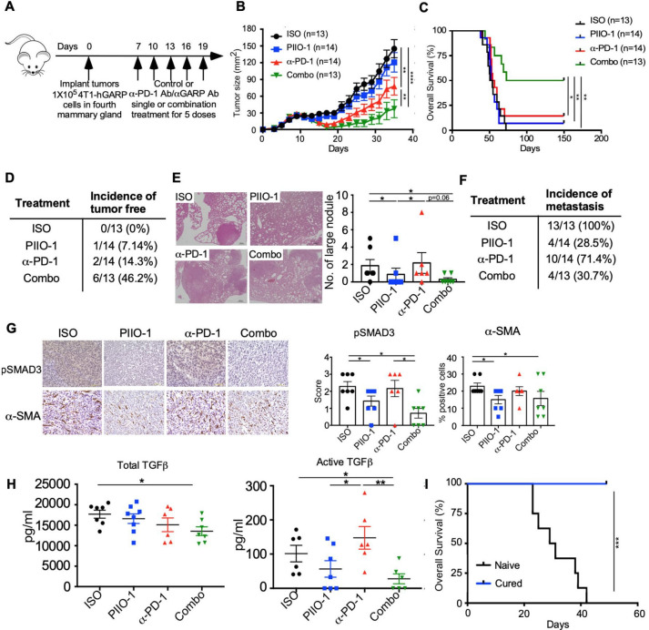
Figure 3.
PIIO-1 enhanced antitumor efficacy of anti-PD-1 ICB in GARP+ TNBC. (A) Experimental scheme. BALB/c mice were injected with 1×10⁵ 4T1-hGARP cells in the mammary fat pad, followed by i.p. injection of 200 µg/mouse of PIIO-1 and/or 150 µg/mouse anti-PD-1 every 3 days. (B) Primary tumor growth curve. (C) Overall survival of mice. (D) Summary of the incidence of tumor-free mice among groups. (E) Lungs were collected and sectioned at experimental end point, then stained with H&E. Representative images from each group of mice are shown. Scale bar, 20 µm. The numbers of visible lung metastatic nodules are quantified. (F) Summary of the incidence of metastasis among groups. (G) Tumors were collected at end point and stained by IHC for pSMAD3, α-SMA. Representative images of tumor tissues from four groups of mice are shown (left). Scale bar, 50 µm. Quantification of the IHC staining is shown (right). (H) Sera from each mouse was collected at end point. Total and active TGFβ level in the sera were assessed by ELISA. (I) Mice with tumor regression following combination therapy were monitored for 300 days, then rechallenged with 5×10⁵ wild-type 4T1 mammary tumor in contralateral mammary fat pad. Naive BALB/c mice without pre-exposure to tumor were used as control. Shown is the overall survival. Tumor curve analysis was performed using repeated measures 2-way analysis of variance. Overall survival is analyzed by log-rank (Mantel-Cox) test. (E, G) were analyzed by paired t-test according to the tumor collection time points. Other data were analyzed by two-tailed Student’s t-test. B, C were corrected for multiple testing using the Tukey procedure. All data are presented as mean±SEM. *p<0.05, **p<0.005, ***p<0.001, ****p<0.0001.

