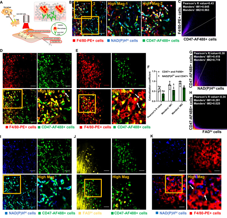
Figure 3.
Imaging of the mechanisms of the CD47-SIRPα pathway in vivo unravels antibody-dependent cellular phagocytosis (ADCP). (A) Schematic diagram of intravital imaging procedure for imaging the mechanisms of the CD47-SIRPα pathway in vivo using triple-negative breast cancer models. (B) Representative images for illustration of the CD47-SIRPα pathway. After injection of anti-CD47-AF488 and F4/80-PE, anti-CD47-AF488 mAb (green) rapidly targeted NAD(P)Hhi cancer cells (blue) indicated by yellow arrows. Engulfment of NAD(P)Hhi and CD47-AF488+ cancer cells by F4/80-PE+ macrophages (red) was well observed (white arrows) presenting ADCP events. (C) Quantitative co-localization analysis using Pearson’s coefficient and Manders’ coefficients further elaborated imaging observation. (D, E) ADCP events occurred as rapid but short wave between F4/80-PE-labeled macrophages (red) and CD47-AF488 mAb-targeted cells (green). CD47 inhibitor-treated cancer cells were first overlapped (yellow arrows) with F4/80+ macrophages and then soon engulfed (white arrows) by the macrophages extensively. (F–J) CD47-AF488 mAb-targeted cells (green) depict metabolic phenotype of strong NAD(P)H expression (blue), (I) whereas no meaningful co-localization with FADhi cells (yellow) (J). Analyses with Peason’s coefficient and Manders’ coefficients corroborated imaging results (F–H). (K) Real-time visualization of phagocytosis events (white arrows) with treatment of CD47 blockade was also achieved using NAD(P)H imaging. Scales, 20 µm. AF, Alexa Flour; CD47, cluster of differentiation 47; FAD, flavin adenine dinucleotide; FADhi, cells high in FAD intensity; mAb, monoclonal antibody; NAD(P)H, reduced nicotinamide adenine dinucleotide (phosphate) hydrogen; NAD(P)Hhi, cells high in NAD(P)H intensity; PE, phycoerythrin; SIRPα, signal regulatory protein α.

