
Figure 1. Global Analysis of HP Secretion.
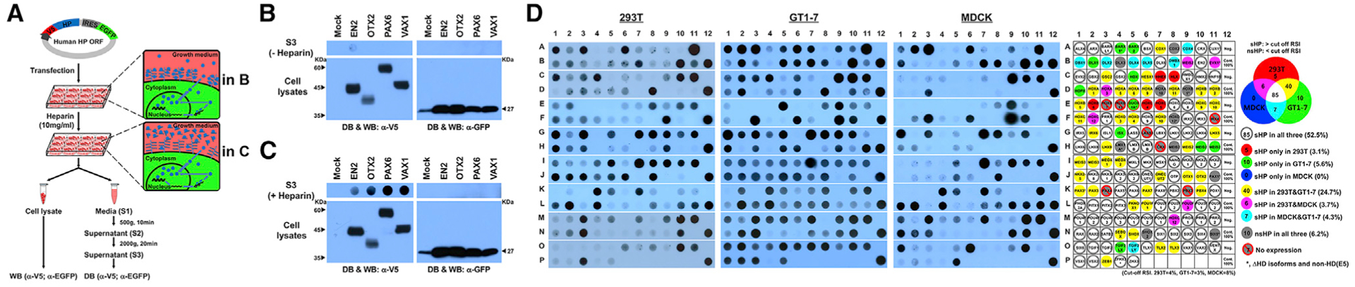
(A) Schematic diagram depicts experimental procedures to detecting secreted HPs.
(B) V5 HPs and EGFP, which are transcribed in same mRNA and translated independently, in the growth media of 293T cells were detected by dot blotting (DB) with anti-V5 and anti-GFP antibodies, respectively. Those proteins expressed in the cells were also detected by western blotting (WB).
(C) Alternatively, the growth media were added with heparin (10 mg/mL) for 3 h, and V5-HPs and EGFP in the growth media were detected.
(D) DB images for V5-HPs in 293T, GT1–7, and MDCK cell growth media treated with heparin (same DB images are provided together with corresponding WB images in Data S1). Secretability of each HP is provided in the virtual DB images in the rightmost column (classification method is provided in Figure S1). Dot colors represent secretion observed in corresponding cell lines.