
Fig. 1.
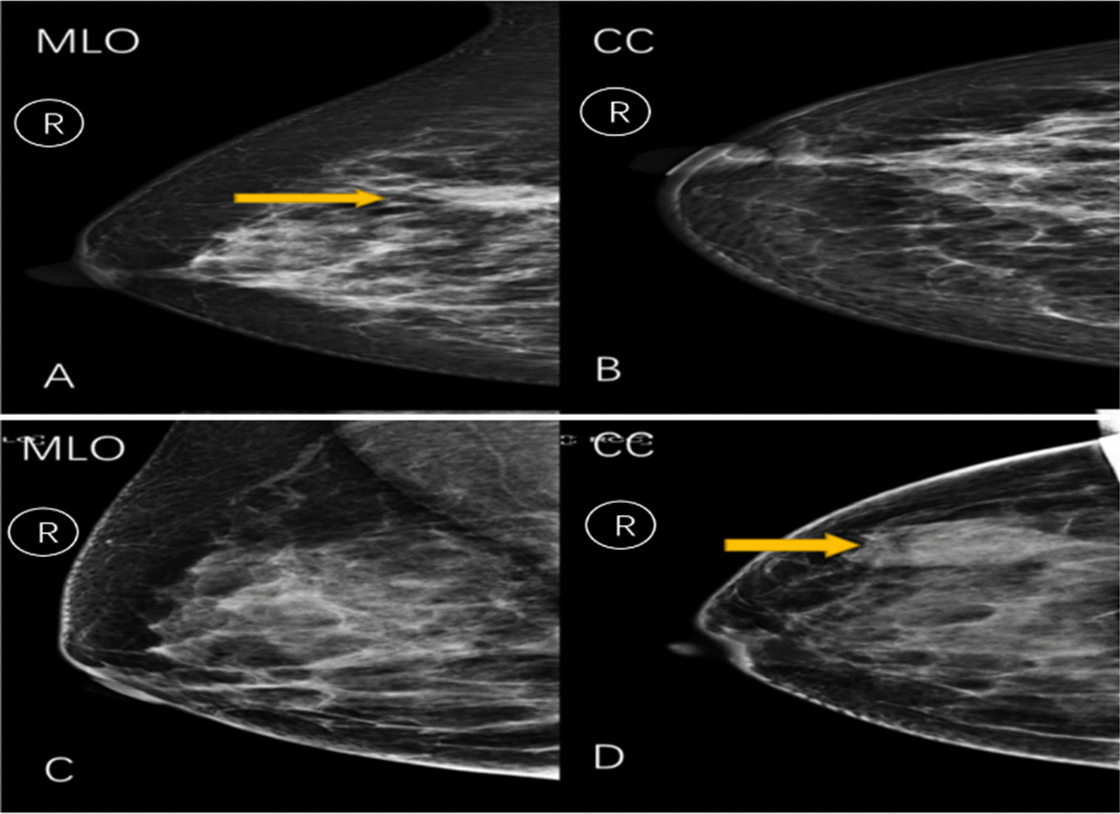
A and B The combination of surgical pathology, ultrasound, and MRI images of a patient (female, 48 years old) determines an irregular mass in the upper inner quadrant of the right breast. A A mass shadow over the right breast is seen in the MLO position in radiography. B No obvious lesion is seen in the CC position; Hence, we have chosen the radiomics features from the MLO. C and D The combination of surgical pathology, ultrasound, and MRI images of a patient (female, 65 years old) shows a well-defined mass in the upper outer quadrant of the right breast. C A mass shadow is seen in the MLO position in radiography. D No obvious lesion is seen in the CC position. C No obvious lesion is observed in the photographic MLO position. D A mass shadow is observed in the right lateral breast in the CC position. Hence, we have chosen the radiomics features from the CC