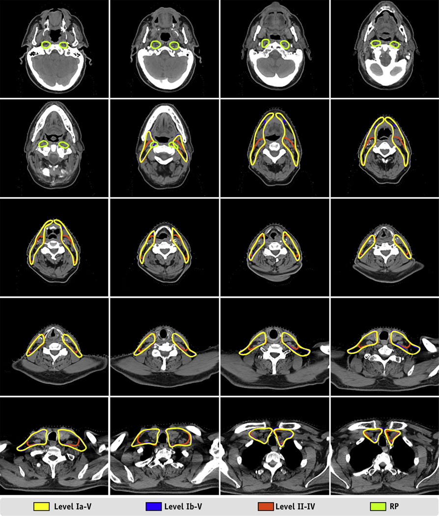
Fig. 4.
Example results from a randomly selected case from our test set. Twenty axial slices from a computed tomography scan of a 57-year-old male patient with base of tongue cancer show the auto-segmented lymph node target volumes. The axial slices are evenly sampled and distributed from the cranial extent of the retropharyngeal lymph nodes to the caudal extent of the level IV lymph node.

