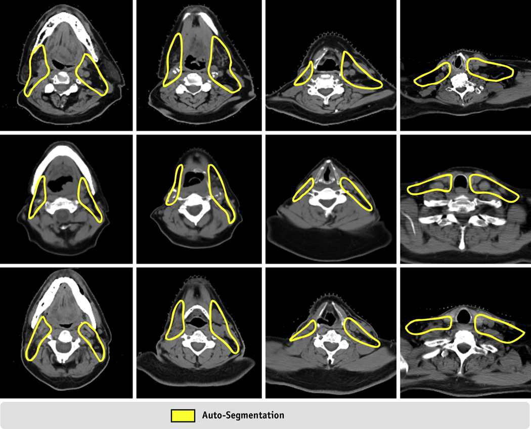
Fig. 5.
Computed tomography images of 3 patients with auto-segmentations requiring minor edits. All 3 patients (1 per row) had their neck dissection before radiation therapy. In these cases, the auto-segmented volumes were undercontoured between lymph node levels II and III as shown in columns 2 and 3. Whereas target volumes for neck lymph node levels Ib-V are shown in this figure, auto-segmentations for levels II-IV and Ia-V were subject to similar undercontouring in these regions. RP node target volumes were unaffected in this clinical presentation.

